

What are anatomical movements?
Anatomical movements are the agreed names for the motions that happen at joints. They are always described from the anatomical position, in which the upper limbs hang beside the body with the palms facing forward. Most movements come in pairs, with one the opposite of the other, such as flexion and extension. (OpenStax Anatomy and Physiology 2e, 9.5: Types of Body Movements.)
TeachMeAnatomy makes the same point: the terms assume that the body starts in the anatomical position, so a movement means the same thing wherever the body happens to be lying or standing. (TeachMeAnatomy: Anatomical Terms of Movement.)
Each pair happens in a typical plane. Flexion and extension take place in the sagittal plane (front to back) around a side-to-side axis; abduction and adduction take place in the frontal (coronal) plane; and rotation takes place in the transverse (horizontal) plane around a vertical axis through the limb. (Kenhub: Types of movements in the human body.)
The shape of a joint limits which movements it allows. A pivot joint, such as the joint between the atlas and the dens of the axis, is uniaxial and allows only rotation. The shoulder and hip are multiaxial ball-and-socket joints: they allow medial and lateral rotation as well as circumduction. (OpenStax Anatomy and Physiology 2e, 9.5: Types of Body Movements.)
The anatomical position and the words for direction: anterior, posterior, medial, lateral, proximal, distal. Directional terms.
Anatomical movements: pairs, planes and examples
| Movement | Opposite | What happens | Plane | Example joint and action |
|---|---|---|---|---|
| Flexion | Extension | Decreases the angle between two parts (bending) | Sagittal | Elbow: bending the arm; hip: raising the thigh forward |
| Hyperextension | None (excess extension) | Extension beyond the normal range | Sagittal | Neck: forced backward beyond its normal range, as in whiplash |
| Lateral flexion | Return to upright | Bending the neck or trunk to one side | Coronal | Vertebral column: leaning sideways |
| Abduction | Adduction | Moves a limb away from the midline | Coronal | Shoulder: raising the arm out to the side; fingers: spreading |
| Medial (internal) rotation | Lateral (external) rotation | Turns the front of a limb toward the midline | Transverse | Hip: turning the toes inward; shoulder: hand toward the opposite hip |
| Rotation (neck and trunk) | Rotation to the other side | Turns the head or trunk around its long axis | Transverse | Atlas around the dens of the axis: shaking the head "no" |
| Circumduction | None | Flexion, abduction, extension and adduction in sequence: a cone | Combines planes | Shoulder or hip: the limb sweeps out a cone |
| Pronation | Supination | Turns the palm backward (down); the radius crosses the ulna | Transverse | Radioulnar joints: turning the palm down |
| Dorsiflexion | Plantar flexion | Lifts the top of the foot toward the shin | Sagittal | Ankle: lifting the front of the foot, heel on the ground |
| Inversion | Eversion | Turns the sole toward the midline | Relative to the median plane | Subtalar and intertarsal joints: the sole faces medially |
| Elevation | Depression | Moves a part upward | Up and down (superior/inferior) | Scapula: shrugging; mandible: closing the mouth |
| Protraction | Retraction | Moves a part forward (scapula: anterolaterally) | Forward and back | Scapula: reaching forward; mandible: jutting the jaw |
| Opposition | Reposition | Brings the thumb tip to a fingertip | Combined movement | First carpometacarpal (saddle) joint of the thumb |
Compiled from OpenStax Anatomy and Physiology 2e §9.5, TeachMeAnatomy and Kenhub. Planes are the usual ones for each movement from the anatomical position; real movements often combine planes.
Flexion and extension (and hyperextension)
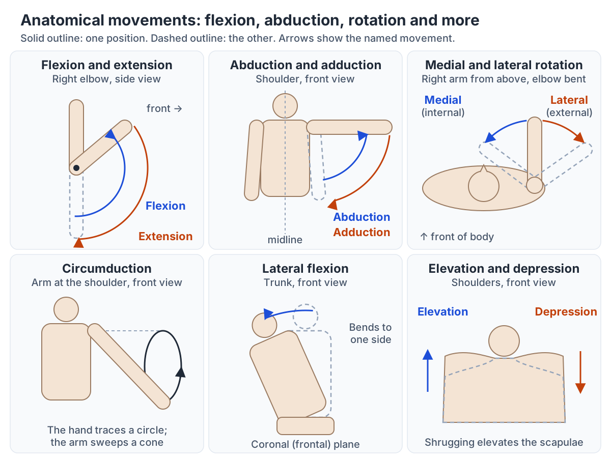
Flexion is a bending movement that decreases the angle between two body parts, such as bending the elbow or knee. Extension is the opposite: a straightening movement that increases the angle between them. Both usually take place in the sagittal plane. Bending the elbow, for example, is flexion: it decreases the angle between the ulna and the humerus. (TeachMeAnatomy: Anatomical Terms of Movement.)
In the neck and trunk, flexion is bending forward and extension is bending backward. In the upper limb every movement forward is flexion and every movement backward is extension, so raising the arm forward at the shoulder is shoulder flexion. Bringing the thigh forward and upward is flexion at the hip, and any backward movement of the thigh is extension. (OpenStax Anatomy and Physiology 2e, 9.5: Types of Body Movements.)
Hyperextension is extension beyond a joint's normal range of motion; hyperflexion is the same for flexion. OpenStax describes hyperextension as the abnormal or excessive extension of a joint, which results in injury, and notes that such injuries are common at hinge joints such as the knee and elbow. In whiplash, when the head is suddenly moved backward and then forward, the neck can be both hyperextended and hyperflexed. (OpenStax Anatomy and Physiology 2e, 9.5: Types of Body Movements.)
Muscles that do these movements work in opposing pairs. The biceps brachii flexes the forearm at the elbow, whereas the triceps brachii extends it; the hamstrings flex the leg at the knee, whereas the quadriceps femoris extend it. (OpenStax Anatomy and Physiology 2e, 11.1: Interactions of Skeletal Muscles, Their Fascicle Arrangement, and Their Lever Systems.)
The sagittal, frontal (coronal) and transverse planes that movements are described in. Anatomical planes.
Lateral flexion of the spine
Lateral flexion is bending the neck or trunk sideways, toward the right or left side. It happens in the coronal (frontal) plane rather than the sagittal plane, and it is a movement of the vertebral column: the neck bends toward one shoulder, or the trunk bends toward one side. (OpenStax Anatomy and Physiology 2e, 9.5: Types of Body Movements.)
Kenhub's movement figures label the same motion lateral flexion of the spine. (Kenhub: Types of movements in the human body.)
What is abduction in anatomy? Abduction and adduction
In anatomy, abduction is movement of a limb away from the midline of the body, as when you raise your arm out to the side. Adduction is the opposite: movement back toward the midline. Both take place in the coronal (frontal) plane. Spreading the fingers or toes apart is also abduction, and bringing them together is adduction. (OpenStax Anatomy and Physiology 2e, 9.5: Types of Body Movements.)
TeachMeAnatomy gives everyday examples: abducting the shoulder raises the arms to the sides, and adducting the hips squeezes the legs together. It adds that for the fingers and toes the midline used is not the body's midline but that of the hand or foot. (TeachMeAnatomy: Anatomical Terms of Movement.)
Kenhub names the reference line more exactly: the fingers move relative to the middle (third) finger and the toes relative to the second toe. At the wrist, moving the hand toward the thumb side is radial deviation and toward the little-finger side ulnar deviation, which OpenStax calls abduction and adduction of the wrist. (Kenhub: Types of movements in the human body.)
The deltoid is the major abductor of the arm at the shoulder. (OpenStax Anatomy and Physiology 2e, 11.5: Muscles of the Pectoral Girdle and Upper Limbs.)
Medial and lateral rotation
Rotation turns a bone around its own long axis. Medial (internal) rotation turns the front of a limb toward the midline of the body, and lateral (external) rotation turns it away from the midline. Rotation happens at ball-and-socket joints such as the shoulder and hip, at pivot joints, and in the vertebral column. (OpenStax Anatomy and Physiology 2e, 9.5: Types of Body Movements.)
TeachMeAnatomy gives two examples: turning a straight leg so that the toes point inward is medial rotation at the hip, and bringing the hand toward the opposite hip with the elbow bent to 90 degrees is internal rotation at the shoulder. (TeachMeAnatomy: Anatomical Terms of Movement.)
In the neck, the atlas (C1) rotates around the dens of the axis (C2), which lets the head turn from side to side, as when shaking the head to say no. (OpenStax Anatomy and Physiology 2e, 9.5: Types of Body Movements.)
At the shoulder, subscapularis is a medial rotator of the arm, while infraspinatus and teres minor rotate it laterally. (TeachMeAnatomy: The Shoulder Joint.)
The ball-and-socket glenohumeral joint and the rotator cuff muscles that rotate the arm. Shoulder anatomy.
What is circumduction?
Circumduction is the movement of a body part in a circular manner. It combines flexion, abduction, extension and adduction one after another at a joint. (OpenStax Anatomy and Physiology 2e, 9.5: Types of Body Movements.)
TeachMeAnatomy notes that circumduction is more accurately a conical movement: the limb sweeps out a cone with its tip at the joint that controls the movement. (TeachMeAnatomy: Anatomical Terms of Movement.)
Sources differ on which joints allow it. Kenhub restricts circumduction to ball-and-socket joints such as the shoulder and hip, while OpenStax also includes biaxial condyloid and saddle joints, such as the saddle joint at the base of the thumb. (Kenhub: Types of movements in the human body.)
Hinge, pivot, condyloid, saddle, plane and ball-and-socket joints and the movements each allows. Types of joints.
Pronation and supination of the forearm
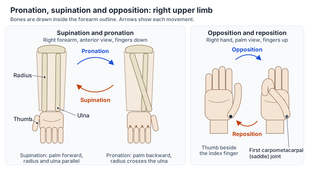
Supination turns the forearm so the palm faces forward (or up, with the hand resting on a table), and pronation turns it so the palm faces backward (or down). They are rotations of the radius around the ulna, at the proximal and distal radioulnar joints, so they happen in the forearm, not at the wrist. (OpenStax Anatomy and Physiology 2e, 9.5: Types of Body Movements.)
In the anatomical position the forearm is supinated and the radius and ulna lie parallel. In pronation the radius rotates at the proximal radioulnar joint and crosses over the ulna, so the two bones form an X; supination uncrosses them. (OpenStax Anatomy and Physiology 2e, 9.5: Types of Body Movements.)
Kenhub describes supination as lateral rotation of the radius and pronation as medial rotation of the radius, in the transverse plane. TeachMeAnatomy describes them with the hand resting on a table and the shoulder and elbow kept still: supination turns the palm up and pronation turns it down. (Kenhub: Types of movements in the human body.)
The axis of this rotation runs obliquely down the forearm, from the centre of the radial head at the elbow to the head of the ulna at the wrist, so the radius swings around the lower end of the ulna. (Tatebe M et al. Tilt of the radius from forearm rotational axis reliably predicts rotational improvement after corrective osteotomy for malunited forearm fractures. Nagoya J Med Sci. 2012.)
Pronator teres and pronator quadratus pronate the forearm. (OpenStax Anatomy and Physiology 2e, 11.5: Muscles of the Pectoral Girdle and Upper Limbs.)
Supinator is the prime mover for slow, unresisted supination and supinates the forearm on its own when the elbow is straight; for quick or forceful supination it is joined by the biceps brachii, which is the most powerful supinator when the elbow is flexed. (Kenhub: Supinator muscle.)
The pronators, the supinator and every other flexor and extensor of the forearm. Forearm muscles.
Dorsiflexion vs plantar flexion
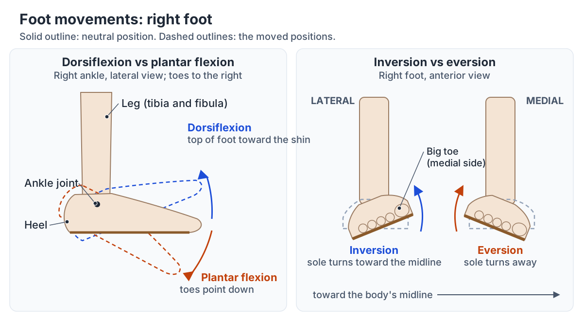
Dorsiflexion is flexion of the top (dorsum) of the foot, which reduces the angle between the foot and the front of the leg. Kenhub's example is lifting the front of the foot while keeping the heel on the ground. Plantar flexion is the opposite: the foot moves down, as when standing on your toes or pointing them. Both happen at the ankle joint. (Kenhub: Types of movements in the human body.)
They are the only movements the ankle joint itself allows. TeachMeAnatomy describes dorsiflexion as flexion at the ankle and plantarflexion as extension at the ankle, so the top-of-foot movement counts as flexion even though the foot moves upward. (TeachMeAnatomy: Anatomical Terms of Movement.)
Different muscle groups do each movement. The anterior compartment muscles of the leg, tibialis anterior, extensor hallucis longus and extensor digitorum longus, dorsiflex the foot. The posterior compartment muscles, gastrocnemius, soleus, plantaris and tibialis posterior, plantar flex it. (TeachMeAnatomy: The Ankle Joint.)
OpenStax agrees that the anterior compartment of the leg is generally responsible for dorsiflexion and the posterior compartment for plantar flexion, and notes that the superficial posterior muscles share the calcaneal (Achilles) tendon on the heel. (OpenStax Anatomy and Physiology 2e, 11.6: Appendicular Muscles of the Pelvic Girdle and Lower Limbs.)
Ankle stability changes with the position. The front of the talus is wider than the back, so in dorsiflexion its broad front part fills the ankle mortise and the joint is more stable; in plantar flexion the narrower back part sits in the mortise and the joint is less stable. (TeachMeAnatomy: The Ankle Joint.)
At the wrist, TeachMeAnatomy notes, the equivalent of dorsiflexion is simply called extension and the equivalent of plantar flexion is palmar flexion. (TeachMeAnatomy: Anatomical Terms of Movement.)
The talocrural hinge joint, its mortise and the ligaments that hold it together. Ankle anatomy.
Dorsiflexion vs plantar flexion
| Feature | Dorsiflexion | Plantar flexion |
|---|---|---|
| What moves | Top of the foot moves toward the shin | Heel lifts or toes point down |
| Joint | Ankle (talocrural) hinge joint | Ankle (talocrural) hinge joint |
| Main muscles | Tibialis anterior, extensor hallucis longus, extensor digitorum longus (anterior compartment) | Gastrocnemius, soleus, plantaris, tibialis posterior (posterior compartment) |
| Tendon at the heel | None | The superficial calf muscles end on the calcaneal (Achilles) tendon |
| Ankle stability | More stable: the wide front of the talus fills the mortise | Less stable: the narrow back of the talus sits in the mortise |
| Everyday example (Kenhub) | Lifting the front of the foot while keeping the heel on the ground | Standing on your toes or pointing them |
| Wrist equivalent | Extension | Palmar flexion |
Based on OpenStax §9.5 and §11.6, TeachMeAnatomy (ankle joint and terms of movement) and Kenhub (everyday examples).
Inversion and eversion of the foot
Inversion of the foot turns the sole toward the midline of the body, so that it faces medially. Eversion of the foot turns the sole away from the midline, so that it faces laterally. These movements help the foot adapt to uneven ground, and the foot can invert further than it can evert. (OpenStax Anatomy and Physiology 2e, 9.5: Types of Body Movements.)
Inversion and eversion do not happen at the ankle joint. OpenStax places them at the plane joints among the tarsal bones of the back of the foot, and TeachMeAnatomy names the subtalar joint, between the talus and the calcaneus, as the chief site for both movements. (TeachMeAnatomy: The Subtalar Joint.)
Pronation and supination are also used for the foot, mostly in clinical, sports and gait contexts, and there they are not the same as eversion and inversion. Because the axis of the subtalar joint is oblique, foot pronation is a combined (triplanar) movement of eversion with abduction and dorsiflexion: the foot rolls inward and the sole turns to face laterally. (Eichelberger P et al. Cranio-caudal and medio-lateral navicular translation are representative surrogate measures of foot function in asymptomatic adults during walking. PLoS One. 2018.)
Supination is the reverse, combining inversion, adduction and plantar flexion, so that the sole faces medially. (Brockett CL, Chapman GJ. Biomechanics of the ankle. Orthop Trauma. 2016.)
Tibialis posterior, the deepest muscle of the posterior compartment, inverts the foot as well as plantar flexing it. (TeachMeAnatomy: Muscles in the Posterior Compartment of the Leg.)
It is usually counted as the main invertor of the foot. (Bizzoca D et al. Polydeoxyribonucleotide in the Treatment of Tendon Disorders, from Basic Science to Clinical Practice: A Systematic Review. Int J Mol Sci. 2023.)
Tibialis anterior also inverts the foot as well as dorsiflexing it, while fibularis (peroneus) tertius, also in the anterior compartment, everts and dorsiflexes it. (TeachMeAnatomy: Muscles in the Anterior Compartment of the Leg.)
The lateral compartment muscles, fibularis longus and fibularis brevis, are the main evertors: their common function is eversion. Fibularis longus also plantar flexes the foot, and TeachMeAnatomy notes that these muscles help to prevent excessive inversion. (TeachMeAnatomy: Muscles in the Lateral Compartment of the Leg.)
Inversion is also how most ankle sprains happen. When the foot rolls inward, the lateral ligaments on the outside of the ankle, the anterior talofibular and calcaneofibular ligaments, are stretched or torn. The less common medial sprain comes from eversion. (OrthoInfo (AAOS): Sprained Ankle.)
Radiopaedia confirms that anterior talofibular ligament injuries typically follow an inversion injury, with or without plantar flexion, and calls it the most commonly injured and weakest ligament of the lateral ankle. (Radiopaedia: Anterior talofibular ligament injury.)
The anterior, lateral and posterior compartments of the leg that move the ankle and foot. Leg muscles.
Elevation, depression, protraction and retraction
Elevation moves a body part upward and depression moves it downward; both describe the scapula and the mandible. Shrugging the shoulders elevates the scapulae, and lowering them depresses the scapulae. In the jaw, elevating the mandible closes the mouth and depressing it opens the mouth. (OpenStax Anatomy and Physiology 2e, 9.5: Types of Body Movements.)
Protraction moves a part forward and retraction moves it back. TeachMeAnatomy describes protraction of the scapula as an anterolateral movement on the chest wall that moves the shoulder forward, and retraction as a posteromedial movement that pulls the shoulder back. (TeachMeAnatomy: Anatomical Terms of Movement.)
The mandible can also protract (push the lower jaw forward) and retract (pull it back), and it moves side to side in lateral and medial excursion. The scapula also rotates: in superior rotation the glenoid cavity turns upward, and in inferior rotation it turns down. (OpenStax Anatomy and Physiology 2e, 9.5: Types of Body Movements.)
Kenhub uses the scapulae as the standard example of protraction and retraction and notes that protraction is anterolateral rather than purely forward, while retraction includes a posteromedial movement. (Kenhub: Types of movements in the human body.)
Opposition and reposition of the thumb
Opposition is the movement that brings the tip of the thumb into contact with the tip of a finger. It happens at the first carpometacarpal joint, a saddle joint at the base of the thumb. Reposition is the return of the thumb to its position beside the index finger. (OpenStax Anatomy and Physiology 2e, 9.5: Types of Body Movements.)
Opposition is a combined movement. OpenStax describes it as flexion plus abduction of the thumb at the carpometacarpal joint, and TeachMeAnatomy explains that opponens pollicis opposes the thumb by medially rotating and flexing the first metacarpal on the trapezium. (TeachMeAnatomy: Muscles of the Hand.)
Kenhub defines opposition as touching the pad of any finger with the thumb of the same hand. (Kenhub: Types of movements in the human body.)
True opposition, in which the first metacarpal rotates on the trapezium so that the thumb pad faces a finger pad, is a feature of the Old World primates: monkeys, apes and humans. New World monkeys such as capuchins have at most a pseudo-opposable thumb. (Aversi-Ferreira RA et al. Anatomical analysis of thumb opponency movement in the capuchin monkey (Sapajus sp). PLoS One. 2014.)
The thumb's saddle joint, the fingers and the muscles that move them. Hand anatomy.
Special cases: the shoulder, thumb, knee and foot
Shoulder: flexion moves the arm forward and extension moves it backward in the sagittal plane, while abduction moves it away from the midline in the coronal plane. Abducting the arm above about 90 degrees is impossible without superior (upward) rotation of the scapula by trapezius and serratus anterior. (TeachMeAnatomy: The Shoulder Joint.)
OpenStax explains the same limit: without superior rotation of the scapula, the arm could not be abducted above shoulder height. (OpenStax Anatomy and Physiology 2e, 9.5: Types of Body Movements.)
The scapula does not wait until 90 degrees, though: after an early setting phase, in which its movement is small and variable, it rotates steadily along with the arm, traditionally about 1 degree for every 2 degrees at the glenohumeral joint (the scapulohumeral rhythm). (Panagiotopoulos AC, Crowther IM. Scapular Dyskinesia, the forgotten culprit of shoulder pain and how to rehabilitate. SICOT J. 2019.)
Thumb: its movements do not follow the fingers' planes. Thumb extension moves it away from the palm within the plane of the palm, and flexion brings it back against the index finger or into the palm. Thumb abduction is a forward movement that brings it perpendicular to the palm, and adduction returns it beside the index finger. (OpenStax Anatomy and Physiology 2e, 9.5: Types of Body Movements.)
Knee: below the knee, flexion moves the leg backward and extension moves it forward, the opposite direction to flexion at the hip or shoulder. OpenStax defines knee flexion as bending the knee to bring the foot toward the back of the thigh. (Kenhub: Types of movements in the human body.)
Foot: the top of the foot is its dorsal surface, so lifting it is called dorsiflexion and pointing the toes is plantar flexion, and turning the sole in or out is inversion or eversion. (TeachMeAnatomy: Anatomical Terms of Movement.)
Frequently asked questions
What is the difference between dorsiflexion and plantar flexion?
They are opposite movements at the ankle. Dorsiflexion lifts the top of the foot toward the shin, as when you lift the front of the foot while keeping the heel on the ground; it is done by the front leg muscles such as tibialis anterior. Plantar flexion points the toes down or lifts the heel, as when you stand on your toes; it is done by the calf muscles, gastrocnemius and soleus.
What is inversion and eversion of the foot?
Inversion turns the sole of the foot inward, toward the midline, so that the sole faces medially. Eversion turns the sole outward, away from the midline. Both happen below the ankle joint, mainly at the subtalar joint between the talus and calcaneus. Most ankle sprains happen when the foot is forced into inversion and the outer ligaments are stretched.
What is abduction in anatomy?
Abduction is moving a body part away from the midline of the body, such as raising your arm or leg out to the side. Its opposite, adduction, brings the part back toward the midline. For the fingers and toes the reference is the middle of the hand or foot, so spreading the fingers is abduction and closing them together is adduction.
What is circumduction?
Circumduction is a circular movement of a limb that combines flexion, abduction, extension and adduction in sequence, so the limb sweeps out a cone with its tip at the joint. When the whole arm circumducts, the tip of that cone is at the shoulder joint. It happens at ball-and-socket joints and, according to OpenStax, at condyloid and saddle joints too.
What is the difference between pronation and supination?
Supination turns the forearm so the palm faces forward or upward, and pronation turns it so the palm faces backward or downward. Both are rotations of the radius around the ulna at the radioulnar joints. In supination the two forearm bones lie parallel; in pronation the radius crosses over the ulna. Supinator and biceps supinate; pronator teres and pronator quadratus pronate.
What is hyperextension?
Hyperextension is extension of a joint beyond its normal range of motion, for example when the neck is forced backward beyond its normal range, as in whiplash. OpenStax describes it as abnormal or excessive extension. Like ordinary extension, it usually happens in the sagittal plane, the front-to-back plane of the body.
What is opposition of the thumb?
Opposition brings the tip of the thumb against the tip of another finger. It is a combined movement at the saddle-shaped first carpometacarpal joint at the base of the thumb, mainly flexion and abduction with medial rotation of the first metacarpal by opponens pollicis. Reposition returns the thumb beside the index finger.
References
- OpenStax. Anatomy and Physiology 2e, §9.5 Types of Body Movements. Rice University, 2022.
- OpenStax. Anatomy and Physiology 2e, §11.1 Interactions of Skeletal Muscles, Their Fascicle Arrangement, and Their Lever Systems. Rice University, 2022.
- OpenStax. Anatomy and Physiology 2e, §11.5 Muscles of the Pectoral Girdle and Upper Limbs. Rice University, 2022.
- OpenStax. Anatomy and Physiology 2e, §11.6 Appendicular Muscles of the Pelvic Girdle and Lower Limbs. Rice University, 2022.
- TeachMeAnatomy. Anatomical Terms of Movement.
- TeachMeAnatomy. The Ankle Joint.
- TeachMeAnatomy. The Subtalar Joint.
- TeachMeAnatomy. Muscles in the Anterior Compartment of the Leg.
- TeachMeAnatomy. Muscles in the Lateral Compartment of the Leg.
- TeachMeAnatomy. The Shoulder Joint.
- TeachMeAnatomy. Muscles of the Hand.
- Kenhub. Types of movements in the human body.
- American Academy of Orthopaedic Surgeons, OrthoInfo. Sprained Ankle.
- Radiopaedia. Anterior talofibular ligament injury.
- TeachMeAnatomy. Muscles in the Posterior Compartment of the Leg.
- Kenhub. Supinator muscle.
- Bizzoca D, Brunetti G, Moretti L, et al. Polydeoxyribonucleotide in the Treatment of Tendon Disorders, from Basic Science to Clinical Practice: A Systematic Review. Int J Mol Sci. 2023;24:4582.
- Aversi-Ferreira RA, Souto Maior R, Aziz A, et al. Anatomical analysis of thumb opponency movement in the capuchin monkey (Sapajus sp). PLoS One. 2014;9:e87288.
- Tatebe M, Shinohara T, Okui N, et al. Tilt of the radius from forearm rotational axis reliably predicts rotational improvement after corrective osteotomy for malunited forearm fractures. Nagoya J Med Sci. 2012;74:167-171.
- Panagiotopoulos AC, Crowther IM. Scapular Dyskinesia, the forgotten culprit of shoulder pain and how to rehabilitate. SICOT J. 2019;5:29.
- Eichelberger P, Pohl J, Jaspers T, et al. Cranio-caudal and medio-lateral navicular translation are representative surrogate measures of foot function in asymptomatic adults during walking. PLoS One. 2018;13:e0208175.
- Brockett CL, Chapman GJ. Biomechanics of the ankle. Orthop Trauma. 2016;30:232-238.
Related anatomy
- Directional termsAnatomical position and directional terms with labelled diagrams: what proximal, distal, medial, lateral, anterior…
- Types of jointsTypes of joints explained: fibrous, cartilaginous and synovial (diarthrosis) joints, a labelled synovial joint diagram…
- Anatomical termsAnatomical terms explained with diagrams: bone markings like tubercle, fossa and condyle, movements like abduction…
- Anatomical planesAnatomical planes explained: midsagittal vs parasagittal, coronal (frontal) and transverse (axial) planes, axes of…
- Ankle jointAnkle anatomy with labelled diagrams and a 3D model: tibia, fibula and talus, the lateral and deltoid ligaments…
- Shoulder jointShoulder anatomy with labelled diagrams and a 3D model: clavicle, scapula and humerus, the glenohumeral, AC and SC…
Explore the whole body in the interactive 3D human anatomy model, or browse the A–Z list of human structures.