

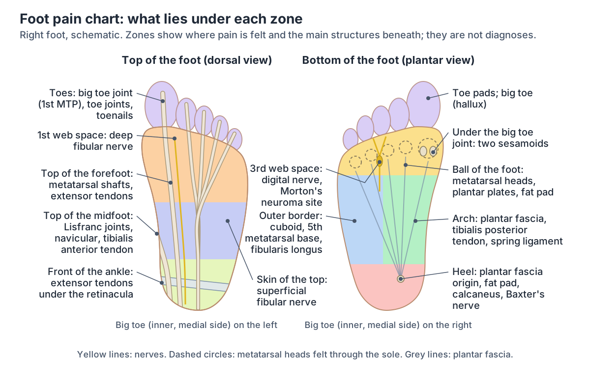
How to read a foot pain chart
Foot pain can be felt on the top, the bottom or the sides of the foot, in the heel, the toes, the arch or anywhere between them. Cleveland Clinic's guide groups the common causes the same way: top of the foot, bottom of the foot (arch and ball), side of the foot, heel, toes, and nerve pain. (Cleveland Clinic: Foot Pain.)
Anatomists divide the foot into three regions. The hindfoot is the talus and calcaneus (heel bone); the midfoot is the navicular, the cuboid and the three cuneiforms; and the forefoot is the metatarsals and the phalanges of the toes. The top of the foot is the dorsal surface and the bottom is the plantar surface (the sole). The inner side, on the big toe side, is medial, and the outer side, on the little toe side, is lateral. (TeachMeAnatomy: Bones of the Foot: Tarsals, Metatarsals and Phalanges.)
A chart works because each region holds different structures. Hospital for Special Surgery (HSS) sorts foot pain into pain on the top of the foot, at the base of the big toe, on the outside and inside of the foot, at the back of the heel (the Achilles tendon), and on the bottom of the foot, which it splits again into the ball (forefoot), the arch (midfoot) and the heel (hindfoot). (Hospital for Special Surgery: Foot Pain Causes, Conditions and Treatments.)
The map is not one-to-one. One structure can hurt in more than one zone: the NHS describes plantar fasciitis pain as felt on the bottom of the foot around both the heel and the arch. (NHS: Plantar fasciitis.)
One zone can also contain several structures that hurt in similar ways, so the NHS foot pain pages list only possible causes for each area and advise not to try to diagnose the cause yourself. Use the chart to learn what lies under each spot. (NHS: Pain in the bottom of the foot.)
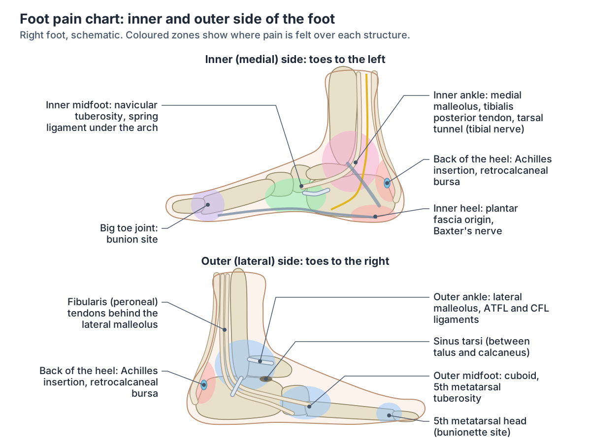
Foot pain chart: zones, the structures under them and linked conditions
| Zone | Main structures there | Conditions linked with that anatomy |
|---|---|---|
| Top of the foot | Extensor tendons, tibialis anterior tendon, metatarsal shafts, midfoot (Lisfranc) joints, navicular | Extensor or tibialis anterior tendonitis, metatarsal or navicular stress fracture, Lisfranc injury, midfoot arthritis, gout |
| Ball of the foot | Metatarsal heads, fat pad, plantar plates, sesamoids, common plantar digital nerves | Metatarsalgia, Morton's neuroma, sesamoiditis, plantar plate tear, Freiberg disease, stress fracture |
| Toes | Big toe (first MTP) joint, plantar plate, smaller toe joints, nail folds | Bunion, hallux rigidus, gout, turf toe, hammer toe, ingrown toenail, fracture |
| Arch | Plantar fascia, tibialis posterior tendon, spring ligament, navicular | Plantar fasciitis, posterior tibial tendon dysfunction, flat feet, tarsal tunnel syndrome (radiating) |
| Heel | Calcaneus, plantar fascia origin, heel fat pad, Achilles insertion, retrocalcaneal bursa, Baxter's nerve | Plantar fasciitis, fat pad syndrome, stress fracture, insertional Achilles tendinopathy, bursitis, Haglund's deformity, Sever's disease (children), Baxter neuropathy |
| Inner side | Medial malleolus, tarsal tunnel (tibial nerve), tibialis posterior tendon, deltoid ligament, first MTP joint | Tarsal tunnel syndrome, posterior tibial tendon dysfunction, medial (eversion) sprain, bunion |
| Outer side | Lateral ligaments (ATFL, CFL), fibularis tendons, sinus tarsi, cuboid, base and head of the fifth metatarsal | Inversion ankle sprain, peroneal tendon disorders, sinus tarsi syndrome, cuboid syndrome, fifth metatarsal fracture, bunionette |
Summary of the zone tables below, each with its sources (NHS, AAOS OrthoInfo, Cleveland Clinic, HSS and peer-reviewed reviews). Educational only: location alone does not identify the cause of foot pain.
Why foot pain is felt where it is
Pain from bones, tendons, ligaments, muscles and skin is somatic pain. It is usually centred on the area of injury or inflammation, which is why a strained tendon or a stress fracture tends to hurt over the structure itself, although deep somatic pain, such as bone pain, can feel more spread out. Nerve (neuropathic) pain is different: it can radiate across an area of the body. (Cleveland Clinic: Somatic Pain.)
Nerve pain can arise anywhere along the nervous system, from the peripheral nerves of the foot to the spinal cord. People describe it as stabbing, shooting or burning, like an electric shock, and it may come with tingling, numbness or a pins and needles feeling. (Cleveland Clinic: Neuropathic Pain.)
Each part of the foot's skin has its own nerve. The superficial fibular nerve supplies most of the top of the foot; the deep fibular nerve supplies the web space between the first and second toes; the saphenous nerve supplies the inner (medial) margin and the sural nerve the outer (lateral) margin of the hindfoot and midfoot. On the sole, the medial plantar nerve supplies the medial two thirds, the lateral plantar nerve the lateral third and the medial calcaneal nerve the inner heel. (TeachMeAnatomy: The Cutaneous Innervation of the Lower Limb.)
Foot symptoms can also start in the lower back. On a dermatome map, the L5 nerve root covers the top and the sole of the foot, the lateral (outer) side of the big toe and toes 2 to 4, while S1 covers the lateral margin of the foot, the heel and the little toe. (Kenhub: Dermatomes.)
The sciatic nerve runs from the lower back to the feet, and the symptoms of sciatica usually affect the buttock and the back of one leg, often including the foot and toes. (NHS: Sciatica.)
Damage to many nerves at once (polyneuropathy) affects the longest nerves first, so its symptoms typically begin in the feet. (NHS: Peripheral neuropathy.)
The L4, L5 and S1 skin bands of the foot and the rest of the dermatome map. Dermatomes.
Top of foot pain: the dorsum, extensor tendons and midfoot
Pain on the top of the foot is felt over the dorsum. Here the tendons of tibialis anterior, extensor hallucis longus and extensor digitorum longus run from the front of the ankle onto the foot: tibialis anterior to the medial cuneiform and the base of the first metatarsal, extensor hallucis longus to the big toe and extensor digitorum longus to the lateral four toes. (TeachMeAnatomy: Muscles in the Anterior Compartment of the Leg.)
Extensor tendonitis is irritation of the tendons that run along the top of the foot. Tendonitis in the foot causes pain along the length of the tendon or where it attaches to bone. (Cleveland Clinic: Foot Tendonitis.)
HSS links each part of the top of the foot with different structures. Pain near the ankle may come from a stress fracture of the navicular; pain after an injury may be a Lisfranc injury of the strong midfoot ligament that holds up the arch; pain over the middle of the foot may come from arthritis of the small midfoot joints or tendonitis of the tibialis anterior tendon; and pain on top of the forefoot over the big toe is often hallux rigidus, arthritis of the big toe joint. (Hospital for Special Surgery: Foot Pain Causes, Conditions and Treatments.)
The metatarsals are the commonest site of stress fractures in the foot. The second and third metatarsals, thinner than the first, are most often affected and cause pain through the middle of the foot. The pain usually builds up gradually and gets worse during weight-bearing activity such as walking, sometimes with swelling on the top of the foot. Navicular stress fractures cause pain that is hard to pinpoint and is worse with weight-bearing. (AAOS OrthoInfo: Stress Fractures of the Foot and Ankle.)
In a Lisfranc injury, bones of the midfoot are broken or the ligaments that support it are torn. The midfoot is where a cluster of small bones forms the arch on the top of the foot. A simple Lisfranc injury can be mistaken for a sprain, but it is a serious injury. (AAOS OrthoInfo: Lisfranc (Midfoot) Injury.)
The NHS lists the possible causes of pain in the top of the foot as a fracture, a sprain or strain, tendonitis or osteoarthritis, gout (which usually starts near the bottom of the big toe), and sciatica, which causes tingling, numbness or pain in the upper foot that may stretch up the back of the leg. (NHS: Pain in the top of the foot.)
See a GP if pain on the top of the foot stops normal activities, keeps coming back or has not improved after 2 weeks. Contact NHS 111 if you cannot walk, the foot has changed shape, you heard a snap at the time of an injury, or the foot is hot and swollen. (NHS: Pain in the top of the foot.)
The navicular, cuneiforms and metatarsals under the top of the foot, labelled from above. Foot bones.
Top of foot pain: structures and linked conditions
| Structure | What can hurt there | Sources |
|---|---|---|
| Extensor hallucis longus and extensor digitorum longus tendons | Extensor tendonitis: pain along the tendon on top of the foot | Cleveland Clinic (foot tendonitis); NHS (top of foot) |
| Tibialis anterior tendon (to medial cuneiform and first metatarsal base) | Tibialis anterior tendonitis: pain on top of the midfoot | HSS; TeachMeAnatomy |
| Second and third metatarsal shafts | Stress fracture: gradual pain, worse with weight-bearing, swelling on top of the foot | AAOS (stress fractures); Palka 2025 |
| Navicular | Stress fracture: pain near the ankle, hard to pinpoint | HSS; AAOS (stress fractures) |
| Tarsometatarsal (Lisfranc) joints and ligaments | Lisfranc injury after a twist or fall; later midfoot arthritis | AAOS (Lisfranc); HSS |
| Small midfoot joints | Midfoot arthritis | HSS; AAOS (arthritis of the foot and ankle) |
| Big toe joint (top) | Hallux rigidus; gout | HSS; NHS (top of foot) |
| Skin nerves: superficial fibular, deep fibular (first web space); L5 root | Tingling, numbness or pain (for example sciatica) | TeachMeAnatomy; Kenhub; NHS (top of foot) |
Ball of foot pain (metatarsalgia)
Pain in the ball of the foot is called metatarsalgia. Its possible causes include exercise such as running, a foot shape that puts extra pressure on the ball, hard or cracked skin, a verruca, Morton's neuroma, bursitis and bunions. (NHS: Pain in the ball of the foot.)
The ball of the foot (forefoot) is the area of the sole between the arch and the toes. Metatarsalgia pain usually centres under one or more of the five metatarsal heads, the ends of the metatarsal bones at the bases of the toes. It can be sharp, shooting, aching or burning and may come with numbness or tingling in the toes. (Cleveland Clinic: Metatarsalgia.)
A 2019 review divides metatarsalgia into primary forms, caused by mechanical overload such as metatarsal malalignment or thinning of the plantar fat pad, and secondary forms caused by other conditions such as Morton neuroma, Köhler disease II (bone necrosis in a metatarsal head) or rheumatoid arthritis. During walking, load is particularly high under the head of the second metatarsal. (Gutteck N, Schilde S, Delank KS. Pain on the plantar surface of the foot. Dtsch Arztebl Int. 2019.)
Morton's neuroma is a thickening of the tissue around a small nerve to the toes, not a true tumour. The common digital plantar nerves pass under the transverse metatarsal ligaments that link the metatarsal heads. The thickening most often develops in the third web space, between the third and fourth toes, and less often in the second web space. (AAOS OrthoInfo: Morton's Neuroma.)
Two sesamoid bones lie under the big toe joint, attached to the big toe flexor tendons, where they act like pulleys and help bear weight. Irritation or inflammation of the tendons around them is sesamoiditis, common in ballet dancers, runners and baseball catchers, and the sesamoids can also fracture from an injury or repeated stress. (AAOS OrthoInfo: Sesamoiditis and Sesamoid Fracture.)
An imaging review adds other structures of the ball: a tear of the plantar plate of the second toe joint (second ray syndrome); Freiberg disease, a subchondral fracture usually of the second metatarsal head in young people; inflamed bursae between the metatarsal heads; and stress fractures, most often of the second and third metatarsals. (Palka O et al. Radiological approach to metatarsalgia in current practice: an educational review. Insights Imaging. 2025.)
The NHS advises seeing a GP if pain in the ball of the foot has not improved after 2 weeks or comes with tingling or loss of sensation, and contacting NHS 111 if you cannot walk, the foot has changed shape, or it is hot, swollen and painful. (NHS: Pain in the ball of the foot.)
The sole, the ball of the foot and the sesamoids in the full foot anatomy guide. Foot anatomy.
Ball of foot pain: structures and linked conditions
| Structure | What can hurt there | Sources |
|---|---|---|
| Metatarsal heads and plantar fat pad | Metatarsalgia from overload or a thinned fat pad | Gutteck 2019; Cleveland Clinic (metatarsalgia) |
| Common plantar digital nerve, usually in the third web space | Morton's neuroma: burning or shooting pain, pebble feeling | AAOS (Morton's neuroma); NHS (Morton's neuroma) |
| Two sesamoid bones under the big toe joint | Sesamoiditis; sesamoid fracture | AAOS (sesamoiditis); Palka 2025 |
| Plantar plate of the second toe joint | Plantar plate tear (second ray syndrome), toe drifting or clawing | Palka 2025; Gutteck 2019 |
| Second metatarsal head | Freiberg disease in young people (bone necrosis of the head) | Palka 2025; Gutteck 2019 (Köhler disease II) |
| Intermetatarsal bursae | Bursitis between the metatarsal heads | Palka 2025; NHS (ball of foot) |
| Skin of the sole | Hard skin, corns, calluses, verruca | NHS (ball and bottom of foot) |
Toe pain: the big toe joint, the smaller toes and the nails
The big toe joint is the metatarsophalangeal (MTP) joint, where the first metatarsal meets the first bone of the big toe. A bunion is a painful bony bump that develops on the inside of the foot at this joint and is linked to hallux valgus, in which the big toe drifts towards the smaller toes. Pain can also spread to the other toes as they crowd and as forces in the ball of the foot change. (AAOS OrthoInfo: Bunions.)
HSS lists the main causes of pain at the base of the big toe as gout, which affects this joint more often than any other; hallux rigidus, degenerative arthritis that stiffens the joint; a bunion; and turf toe, a sprain of the joint when the toe bends upwards too far. (Hospital for Special Surgery: Foot Pain Causes, Conditions and Treatments.)
Turf toe is a sprain, partial tear or complete tear of the plantar plate of the first MTP joint, usually caused by forced bending of the big toe upwards (hyperextension). A sesamoid can occasionally fracture at the same time. (Radiopaedia: Turf toe.)
Gout is caused by uric acid crystals deposited in joints, and its first attacks often occur in the joints of the big toe. The affected joint becomes red, swollen, hot and very tender. (AAOS OrthoInfo: Gout.)
Hammer toe affects the second, third or fourth toe, which bends at its middle joint, the proximal interphalangeal (PIP) joint. Pressure from shoes can raise a corn or callus on top of the bent joint or at the tip of the toe. (AAOS OrthoInfo: Hammer Toe.)
The NHS lists the possible causes of toe pain as an ingrown toenail, a bunion, Raynaud's or chilblains (pain, tingling and numbness when cold or stressed, with colour change), a sprained or broken toe, and gout. (NHS: Toe pain.)
An ingrown toenail forms when the corner of the nail grows down into the skin beside it. It usually affects the big toe but can affect any toe. (Cleveland Clinic: Ingrown Toenails.)
Contact NHS 111 if you have badly hurt your big toe, a toe points at an odd angle, you have difficulty moving your toes or you cannot walk, you have severe pain or any tingling or loss of sensation in the foot, or you have toe pain and feel hot, cold or shivery. (NHS: Toe pain.)
Toe pain: structures and linked conditions
| Structure | What can hurt there | Sources |
|---|---|---|
| First metatarsophalangeal (big toe) joint, inner side | Bunion (hallux valgus) | AAOS (bunions); NHS (toe pain) |
| First metatarsophalangeal joint cartilage | Hallux rigidus (arthritis, stiffness) | HSS; Palka 2025 |
| Big toe joint | Gout: sudden, red, hot, swollen joint | AAOS (gout); NHS (gout) |
| Plantar plate of the first MTP joint | Turf toe after forced upward bending | Radiopaedia (turf toe); HSS |
| Middle (PIP) joint of toes 2 to 4 | Hammer toe, with corns over the joint | AAOS (hammer toe); HSS |
| Nail and nail fold | Ingrown toenail, usually the big toe | Cleveland Clinic (ingrown toenails); NHS (toe pain) |
| Phalanges | Fracture after a direct blow, for example stubbing or dropping something | AAOS (toe and forefoot fractures); NHS (toe pain) |
| Small blood vessels of the toes | Raynaud's or chilblains: pain with cold and colour change | NHS (toe pain); Cleveland Clinic (foot pain) |
Foot arch pain: plantar fascia, tibialis posterior and the spring ligament
The plantar fascia is a strong fibrous band that runs from the heel to the ball of the foot and the toes and forms the arch on the bottom of the foot. Plantar fasciitis, inflammation of this band, is the most common cause of heel pain and can be felt in the heel or along the bottom of the foot. (Cleveland Clinic: Plantar Fasciitis.)
A 2019 review describes the plantar fascia, together with the plantar calcaneonavicular (spring) ligament and the long plantar ligament, as the passive tensioning structures of the longitudinal arch. The fascia also gives origin to the short muscles of the sole. (Gutteck N, Schilde S, Delank KS. Pain on the plantar surface of the foot. Dtsch Arztebl Int. 2019.)
The posterior tibial tendon runs behind the medial malleolus (the inside ankle bump) to the bones on the inside of the arch, and its main job is to hold the arch up. The spring ligament, from the calcaneus to the navicular, also supports the arch. When the tendon becomes inflamed or torn, pain is felt in the arch or along the inside of the foot and ankle, sometimes before any change in foot shape. (AAOS OrthoInfo: Progressive Collapsing Foot Deformity (Posterior Tibial Tendon Dysfunction).)
Posterior tibial tendon dysfunction is the leading cause of progressive collapsing foot deformity, formerly called adult-acquired flatfoot or fallen arch. (Cleveland Clinic: Posterior Tibial Tendon Dysfunction.)
In flat feet (pes planus) the arch on the inner side of the sole is flattened and appears to touch the ground. Flat feet do not always hurt, but arches that never develop or that collapse later in life can cause pain. (Cleveland Clinic: Flat Feet (Pes Planus).)
Pain in the arch can also come from nearby. In tarsal tunnel syndrome, pain starts at the inner ankle and spreads into the sole, with burning, tingling or numbness, and it usually gets worse with standing or walking. (Rodríguez-Merchán EC, Moracia-Ochagavía I. Tarsal tunnel syndrome: current rationale, indications and results. EFORT Open Rev. 2021.)
HSS adds that problems in the forefoot or hindfoot can cause secondary symptoms in the arch: a Morton's neuroma, for example, can feel like walking on a pebble and cause discomfort felt in the arch. (Hospital for Special Surgery: Foot Pain Causes, Conditions and Treatments.)
See a GP if pain in the bottom of the foot keeps coming back, has not improved after 2 weeks, or comes with tingling, loss of sensation or weak feet. Contact NHS 111 if the pain is severe, you cannot walk or the foot has changed shape. (NHS: Pain in the bottom of the foot.)
The plantar layers that lie between the plantar fascia and the bones of the arch. Foot muscles.
Foot arch pain: structures and linked conditions
| Structure | What can hurt there | Sources |
|---|---|---|
| Plantar fascia (plantar aponeurosis) | Plantar fasciitis: pain around the heel and arch | NHS (plantar fasciitis); Cleveland Clinic (plantar fasciitis) |
| Tibialis posterior tendon (to the navicular and medial tarsal bones) | Posterior tibial tendon dysfunction: pain in the arch and inner ankle | AAOS (PCFD); Cleveland Clinic (PTTD) |
| Spring (plantar calcaneonavicular) ligament | Stretched or torn as the arch collapses | AAOS (PCFD); Gutteck 2019 |
| Shape of the medial longitudinal arch | Flat feet (fallen arches), which may or may not hurt | NHS (bottom of foot); Cleveland Clinic (flat feet) |
| Navicular | Stress fracture: pain hard to pinpoint | AAOS (stress fractures); HSS |
| Tibial and plantar nerves | Tarsal tunnel syndrome radiating into the sole | Rodríguez-Merchán 2021; Cleveland Clinic (tarsal tunnel) |
Heel pain: under the heel and at the back of the heel
Heel pain is a very common foot and ankle problem. It can be felt anywhere in the heel, but most often underneath it or at the back of it. (Cleveland Clinic: Heel Pain.)
The heel bone (calcaneus) is the largest bone in the foot. The Achilles tendon attaches to the back of it and the plantar fascia to its underside, and both attachments can become inflamed and cause heel pain. (AAOS OrthoInfo: Heel Pain.)
A 2019 review of plantar foot pain describes the pain of plantar fasciitis in the medial (inner) part of the underside of the heel, typically with the first steps after getting out of bed or after a rest. On examination, clinicians find tenderness at the attachment of the fascia to the calcaneal tubercle, and the condition peaks between 45 and 65 years of age. (Gutteck N, Schilde S, Delank KS. Pain on the plantar surface of the foot. Dtsch Arztebl Int. 2019.)
AAOS gives the most common age for plantar fasciitis as 40 to 60, and notes that most people with a heel spur on an X-ray have no heel pain: a spur is a result of long-standing tension at the fascia's attachment, not the cause of the pain. (AAOS OrthoInfo: Plantar Fasciitis and Bone Spurs.)
Plantar heel pain is an umbrella term. A 2022 scoping review lists plantar fasciopathy, heel fat pad syndrome, nerve irritation, calcaneal stress fracture and lumbar radiculopathy among its causes. In one of the studies it reviewed, heel fat pad syndrome was diagnosed from a heel fat pad thinner than 3 mm on ultrasound together with pain at the centre or edge of the heel that was worse barefoot or after long standing. (Chang AH et al. A scoping review of heel fat pad syndrome. J Foot Ankle Res. 2022.)
Pain behind the heel may come from retrocalcaneal bursitis, inflammation where the Achilles tendon inserts on the heel bone, or from insertional Achilles tendinosis, breakdown of the tendon at its attachment. Under the heel, AAOS also lists a bruised fat pad (stone bruise), a calcaneus stress fracture and nerve entrapment beneath the heel bone. (AAOS OrthoInfo: Heel Pain.)
Cleveland Clinic adds Haglund's deformity, an enlarged bony bump at the back of the heel (a pump bump), and Sever's disease in children, an irritation of the growth plate at the back of the heel that is common in active children between 8 and 14. (Cleveland Clinic: Heel Pain.)
A paediatric review describes Sever's disease (calcaneal apophysitis) as pain where the Achilles tendon inserts into the calcaneal apophysis, the growth centre of the heel, and gives a typical age of 8 to 15 years, since the growth plate does not usually close before 14. (Kothari EA et al. A Review of Pediatric Heel Pain. Cureus. 2023.)
Contact NHS 111 if heel pain is severe after an injury, you heard a snap or popping noise, or you cannot walk or walk on tiptoes. See a GP if heel pain has not improved after 2 weeks, comes with tingling or loss of sensation, or if you have diabetes. (NHS: Heel pain.)
The heel bone in 3D, with the attachments of the Achilles tendon and plantar fascia. Calcaneus.
Heel pain: structures and linked conditions
| Structure | What can hurt there | Sources |
|---|---|---|
| Plantar fascia origin at the medial calcaneal tubercle | Plantar fasciitis: inner underside of the heel, first steps worst | Gutteck 2019; AAOS (plantar fasciitis) |
| Heel fat pad | Fat pad syndrome or bruise: centre of the heel | Chang 2022; AAOS (heel pain) |
| Calcaneus | Stress fracture; fracture after a fall | AAOS (heel pain); Cleveland Clinic (heel pain) |
| Inferior calcaneal (Baxter's) nerve | Baxter neuropathy: plantar medial heel pain | Radiopaedia (Baxter neuropathy); Rodríguez-Merchán 2021 |
| Achilles tendon insertion | Insertional Achilles tendinopathy | AAOS (Achilles tendinitis); AAOS (heel pain) |
| Retrocalcaneal bursa (between tendon and bone) | Retrocalcaneal bursitis | AAOS (heel pain); Cleveland Clinic (heel bursitis) |
| Back of the calcaneus | Haglund's deformity (pump bump) | Cleveland Clinic (heel pain); HSS |
| Calcaneal apophysis (growth plate) | Sever's disease in children of about 8 to 15 | Cleveland Clinic (heel pain); Kothari 2023 |
Foot inside pain: the medial side of the foot and ankle
The tarsal tunnel lies on the posteromedial side of the ankle, around the back of the medial malleolus. Its roof is the flexor retinaculum, a band stretched from the medial malleolus to the medial tubercle of the calcaneus. Through it pass the tibialis posterior tendon, the flexor digitorum longus tendon, the posterior tibial artery and vein, the tibial nerve and the flexor hallucis longus tendon. (TeachMeAnatomy: The Tarsal Tunnel.)
With posterior tibial tendon problems, swelling and pain are usually found just behind and below the inside ankle bump, and the arch itself may hurt. As the arch collapses, the heel bone can shift outwards and press on the outer ankle bone, so pain may later appear on the outside of the ankle as well. (AAOS OrthoInfo: Progressive Collapsing Foot Deformity (Posterior Tibial Tendon Dysfunction).)
Tarsal tunnel syndrome is compression of the tibial nerve in the tarsal tunnel. It usually causes pain on the inside of the ankle or the bottom of the foot, with burning or tingling in the sole and toes. (Cleveland Clinic: Tarsal Tunnel Syndrome.)
Pain on the inner heel, in the medial part of the underside of the heel, is typical of plantar fasciitis. (Gutteck N, Schilde S, Delank KS. Pain on the plantar surface of the foot. Dtsch Arztebl Int. 2019.)
Baxter neuropathy, compression of the inferior calcaneal nerve (Baxter's nerve), causes heel pain with the most tenderness on the plantar medial side of the foot, just in front of the medial side of the calcaneus. (Radiopaedia: Baxter neuropathy.)
Further forward, the bunion of hallux valgus forms on the inside of the foot at the big toe joint. (AAOS OrthoInfo: Bunions.)
Ankle sprains on the inner side are less common than those on the outer side. A medial ankle sprain is an eversion injury, with the foot turning out, that stretches or tears the deltoid ligament on the inside of the ankle. (AAOS OrthoInfo: Sprained Ankle.)
For pain around the ankle, the NHS advises contacting 111 if the pain is severe, you cannot walk, the ankle or foot is at an odd angle or you heard a snap at the time of injury, and seeing a GP if there is tingling or loss of feeling in the foot. (NHS: Ankle pain.)
The medial malleolus, the deltoid ligament and the tendons behind the inner ankle. Ankle anatomy.
Foot inside pain: structures and linked conditions
| Structure | What can hurt there | Sources |
|---|---|---|
| Tibialis posterior tendon behind and below the medial malleolus | Posterior tibial tendon dysfunction | AAOS (PCFD); Cleveland Clinic (PTTD) |
| Tibial nerve in the tarsal tunnel | Tarsal tunnel syndrome: inner ankle pain spreading into the sole | Rodríguez-Merchán 2021; Cleveland Clinic (tarsal tunnel) |
| Plantar fascia origin and Baxter's nerve (inner heel) | Plantar fasciitis; Baxter neuropathy | Gutteck 2019; Radiopaedia (Baxter neuropathy) |
| Deltoid ligament | Medial ankle sprain (eversion), less common | AAOS (sprained ankle); AAOS (PCFD) |
| First MTP joint, inner side | Bunion | AAOS (bunions); HSS |
Pain on the outside of the foot (lateral foot pain)
Most ankle sprains are inversion injuries, in which the foot turns inwards and stretches or tears the anterior talofibular ligament (ATFL) and the calcaneofibular ligament (CFL), the lateral ligaments on the outside of the ankle. (AAOS OrthoInfo: Sprained Ankle.)
Just behind the lateral malleolus, the tendons of fibularis (peroneus) brevis and longus share a groove under a retinaculum. Brevis then runs to the base of the fifth metatarsal and longus turns under the cuboid into the sole. Problems of these tendons (tendinopathy, slipping out of the groove, and splits or tears) are an under-recognised cause of pain on the outer side of the hindfoot and can be hard to tell apart from a sprained lateral ligament. (Davda K et al. Peroneal tendon disorders. EFORT Open Rev. 2017.)
Peroneal tendonitis causes pain along the outside of the foot and ankle and can follow a sudden ankle sprain as well as overuse. (Cleveland Clinic: Peroneal Tendonitis.)
Cuboid syndrome is thought to arise from a subtle disruption of the calcaneocuboid joint, between the heel bone and the cuboid, and is considered a common source of lateral midfoot pain in athletes. Its symptoms resemble a ligament sprain, it can follow an ankle sprain, and there is no definitive test for it. (Durall CJ. Examination and treatment of cuboid syndrome: a literature review. Sports Health. 2011.)
Fibularis brevis attaches to the tuberosity at the base of the fifth metatarsal, fibularis tertius just beyond it, and the lateral band of the plantar fascia to the base. Tuberosity avulsion fractures (zone 1) usually happen in inversion injuries, from the pull of fibularis brevis and the lateral band of the plantar fascia. A Jones fracture (zone 2) lies a little further along, at the junction of the base and the shaft, where the blood supply is poorer. (Chloros GD et al. Fifth metatarsal fractures: an update on management, complications, and outcomes. EFORT Open Rev. 2022.)
Stress fractures of the fourth and fifth metatarsals cause pain on the outside of the foot, and those at the base of the fifth metatarsal heal slowly because this area has a poor blood supply. (AAOS OrthoInfo: Stress Fractures of the Foot and Ankle.)
The sinus tarsi is the small space between the talus and the calcaneus, at the lower outer side of the ankle. In sinus tarsi syndrome, irritated tissue there causes painful swelling on the lower outside part of the ankle, and ankle sprains are the most common cause. (Cleveland Clinic: Sinus Tarsi Syndrome.)
A bunionette, or tailor's bunion, is the same kind of bump as a bunion but on the outside of the foot, near the base of the little toe. (AAOS OrthoInfo: Bunions.)
After a twisting injury, contact NHS 111 if you cannot walk, the ankle or foot has changed shape, you heard a snap, grinding or popping noise, or you feel faint, dizzy or sick from the pain: these can be signs of a broken ankle. (NHS: Ankle pain.)
The bone whose tuberosity you can feel on the outer edge of the foot, in 3D. Fifth metatarsal.
Outer side foot pain: structures and linked conditions
| Structure | What can hurt there | Sources |
|---|---|---|
| Anterior talofibular and calcaneofibular ligaments | Inversion ankle sprain | AAOS (sprained ankle); Radiopaedia (ankle sprain) |
| Fibularis (peroneus) brevis and longus tendons behind the lateral malleolus | Peroneal tendinopathy, subluxation, splits and tears | Davda 2017; Cleveland Clinic (peroneal tendonitis) |
| Sinus tarsi (between talus and calcaneus) | Sinus tarsi syndrome after sprains | Radiopaedia (sinus tarsi); Cleveland Clinic (sinus tarsi) |
| Calcaneocuboid joint and cuboid | Cuboid syndrome | Durall 2011; Cleveland Clinic (cuboid syndrome) |
| Base of the fifth metatarsal | Tuberosity avulsion fracture; Jones fracture; stress fracture | Chloros 2022; AAOS (stress fractures); Cleveland Clinic (Jones fracture) |
| Fourth and fifth metatarsal shafts | Stress fracture with pain on the outside of the foot | AAOS (stress fractures); HSS |
| Fifth metatarsal head | Bunionette (tailor's bunion) | AAOS (bunions); Cleveland Clinic (bunions) |
| Sural nerve; S1 root | Tingling or pain along the outer edge of the foot | TeachMeAnatomy; Kenhub |
Nerve pain in the foot: tarsal tunnel, Morton's neuroma and neuropathy
Nerve pain in the foot can feel like burning, tingling or an electric shock. Cleveland Clinic lists burning feet syndrome, Baxter's neuropathy, tarsal tunnel syndrome, peripheral neuropathy, sciatica and diabetes-related neuropathy among its causes. (Cleveland Clinic: Foot Pain.)
In tarsal tunnel syndrome the tibial nerve and its branches are compressed in the tarsal tunnel, beneath the flexor retinaculum. The tibial nerve gives rise to the medial and lateral plantar nerves that supply the sole, and the syndrome is often underdiagnosed or mistaken for plantar fasciitis. (Rodríguez-Merchán EC, Moracia-Ochagavía I. Tarsal tunnel syndrome: current rationale, indications and results. EFORT Open Rev. 2021.)
The first branch of the lateral plantar nerve is known as Baxter's nerve and supplies the abductor digiti minimi muscle. It is usually compressed between the deep fascia of the abductor hallucis muscle and the quadratus plantae muscle, and this compression can cause heel pain with or without plantar fasciitis. (Rodríguez-Merchán EC, Moracia-Ochagavía I. Tarsal tunnel syndrome: current rationale, indications and results. EFORT Open Rev. 2021.)
Morton's neuroma is fibrosis in and around a common plantar digital nerve where it divides between two metatarsal heads. It typically affects middle-aged women and causes sharp pain between the metatarsal heads, sometimes like an electric shock, which eases when the shoe is taken off. (Palka O et al. Radiological approach to metatarsalgia in current practice: an educational review. Insights Imaging. 2025.)
The NHS describes the pain of Morton's neuroma as shooting, stabbing or burning between the third and fourth toes, often with a feeling of a pebble under the foot and sometimes with tingling or numbness. (NHS: Morton's neuroma.)
Peripheral neuropathy is damage to the nerves of the hands, feet and limbs. Its main symptoms are numbness and tingling in the feet or hands, burning, stabbing or shooting pain, loss of balance and muscle weakness, especially in the feet. In the UK, diabetes is the most common cause. (NHS: Peripheral neuropathy.)
Cleveland Clinic also names diabetes as the most common cause overall, often affecting the nerves in the feet first. (Cleveland Clinic: Peripheral Neuropathy.)
A review of plantar foot pain describes the pain of polyneuropathy in people with diabetes as burning and usually independent of loading. (Gutteck N, Schilde S, Delank KS. Pain on the plantar surface of the foot. Dtsch Arztebl Int. 2019.)
Nerve root problems can imitate foot nerve entrapment: an S1 radiculopathy can give patchy symptoms in the sole, usually on one side, while symptoms in both feet raise the question of polyneuropathy. (Rodríguez-Merchán EC, Moracia-Ochagavía I. Tarsal tunnel syndrome: current rationale, indications and results. EFORT Open Rev. 2021.)
Severe pain, numbness or weakness that is quickly getting worse needs emergency care, because it can be a sign of nerve damage. (Cleveland Clinic: Neuropathic Pain.)
The tibial, fibular, sural and saphenous nerves from the knee to the foot. Nerves of the leg.
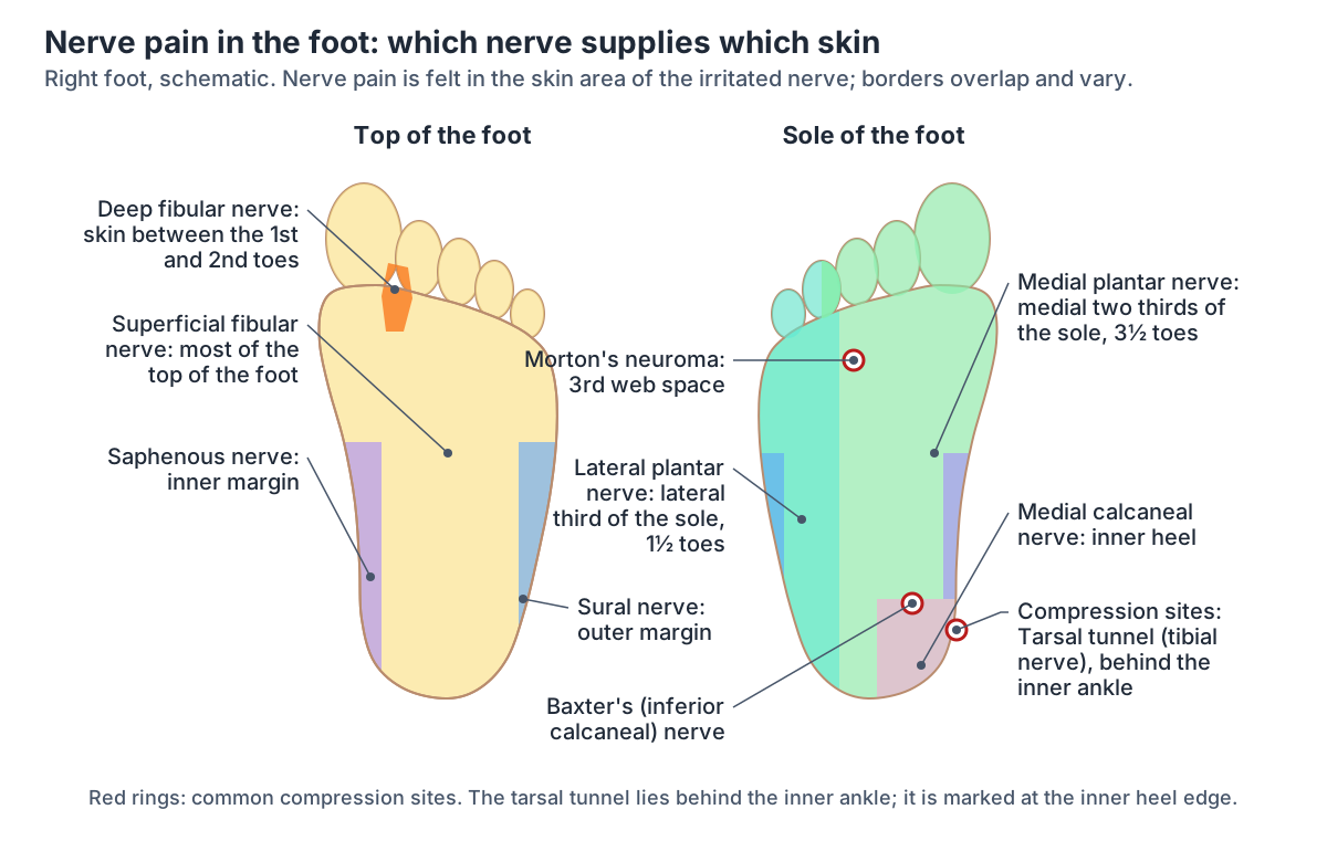
Nerve pain in the foot: which nerve, where it is felt
| Nerve | Where symptoms are felt | Linked condition | Sources |
|---|---|---|---|
| Tibial nerve in the tarsal tunnel | Inner ankle, spreading into the sole and toes | Tarsal tunnel syndrome | Rodríguez-Merchán 2021; Cleveland Clinic |
| Inferior calcaneal (Baxter's) nerve | Plantar medial heel | Baxter neuropathy | Radiopaedia; Rodríguez-Merchán 2021 |
| Common plantar digital nerve (usually third web space) | Ball of the foot and between the third and fourth toes | Morton's neuroma | AAOS; NHS; Palka 2025 |
| Many long nerves (polyneuropathy) | Both feet first, then higher | Peripheral neuropathy, most often from diabetes | NHS; Cleveland Clinic |
| L5 and S1 nerve roots (sciatic nerve) | L5: top and sole of the foot; S1: outer edge and heel | Sciatica, radiculopathy | Kenhub; NHS (sciatica) |
Finding the painful spots in the 3D model
Several landmarks in the model match zones on the chart. The calcaneus forms the heel, and the Achilles tendon attaches to the calcaneal tuberosity at its back. The navicular sits on the inner side of the midfoot, with a tuberosity for part of the tibialis posterior tendon, and the cuboid on the outer side has a groove on its underside for the fibularis longus tendon. (TeachMeAnatomy: Bones of the Foot: Tarsals, Metatarsals and Phalanges.)
The two sesamoid bones are always present under the head of the first metatarsal, and the tuberosity at the base of the fifth metatarsal can be seen and felt on the outer border of the foot, where fibularis brevis inserts. (Kenhub: Metatarsal bones.)
The 3D view above is the BodyParts3D adult male model, showing the outer (lateral) side of the left foot; turn it to see the inner side and the sole. It highlights the bones of the foot, the long plantar ligament, abductor hallucis, flexor digitorum brevis and abductor digiti minimi. The Achilles (calcaneal) tendon, fibularis brevis, fibularis longus, tibialis posterior and tibialis anterior run up into the calf, so open their own pages to see them. The model has no plantar fascia, nerves, bursae or fat pads; use the three diagrams on this page for those.
The deep calf muscle whose tendon supports the arch, in 3D. Tibialis posterior.
When to see a doctor for foot pain
The NHS advises seeing a GP if foot pain stops you doing normal activities, is getting worse or keeps coming back, has not improved after 2 weeks, or comes with tingling or loss of sensation in the foot, and if you have diabetes and foot pain, because foot problems can be more serious with diabetes. (NHS: Pain in the top of the foot.)
Contact NHS 111 urgently if the pain is severe, you cannot walk, the foot has changed shape or is at an odd angle, you heard a snap, grinding or popping noise at the time of an injury, or you feel faint, dizzy or sick from the pain. A foot that is hot and swollen, or pain with a very high temperature or feeling hot, cold and shivery, can be a sign of infection. (NHS: Pain in the top of the foot.)
For heel pain, the NHS adds sudden sharp pain with a popping or snapping sound (which can mean a heel fracture or a ruptured Achilles tendon), not being able to walk on tiptoes or up stairs, and swelling and bruising in the calf and ankle. (NHS: Heel pain.)
Sudden pain and swelling in a joint together with a very high temperature, or feeling hot, cold or shivery, could mean an infection inside the joint and needs urgent medical help. (NHS: Gout.)
Go to A&E or call 999 if back-related leg and foot symptoms come with sciatica on both sides, severe or worsening weakness or numbness in both legs, numbness around the genitals or anus, or new problems controlling the bladder or bowels. (NHS: Sciatica.)
See a GP early for pain, tingling or loss of sensation in the feet, loss of balance or weakness, or a cut or ulcer on the foot that is not getting better, which can be early signs of peripheral neuropathy. (NHS: Peripheral neuropathy.)
Cleveland Clinic gives similar reasons to book an appointment: foot pain that lasts more than two weeks without improving, sudden or severe pain, pain after an injury, signs of infection such as redness, swelling or fever, and diabetes or another condition that affects blood flow. (Cleveland Clinic: Foot Pain.)
Foot pain chart in the 3D model
- Abductor digiti minimi of foot
- A muscle along the lateral border of the sole of the foot.
- Abductor hallucis
- A muscle along the medial border of the sole of the foot.
- Calcaneus
- The largest tarsal bone, forming the heel.
- Cuboid bone
- A cube-shaped tarsal bone on the lateral side of the foot.
- Distal phalanx of big toe
- The end bone of the big toe (hallux), broad and stout.
- Distal phalanx of fourth toe
- The small end bone of the fourth toe (fourth digit of the foot).
- Distal phalanx of little toe
- The small end bone of the little toe (fifth digit of the foot).
- Distal phalanx of second toe
- The small end bone of the second toe (second digit of the foot).
- Distal phalanx of third toe
- The small end bone of the third toe (third digit of the foot).
- Fifth metatarsal bone
- The metatarsal of the little toe. One of the five long bones of the forefoot.
- First metatarsal bone
- The shortest, thickest metatarsal, supporting the big toe. One of the five long bones of the forefoot.
- Flexor digitorum brevis
- A central muscle of the sole.
- Fourth metatarsal bone
- A lesser metatarsal on the lateral side of the foot. One of the five long bones of the forefoot.
- Intermediate cuneiform bone
- The smallest of the three cuneiform bones.
- Lateral cuneiform bone
- A wedge-shaped bone of the midfoot.
- Long plantar ligament
- The longest ligament of the sole of the foot.
- Medial cuneiform bone
- The largest of the three cuneiform bones.
- Middle phalanx of fourth toe
- The middle phalanx of the fourth toe (fourth digit of the foot).
- Middle phalanx of little toe
- The middle phalanx of the little toe (fifth digit of the foot).
- Middle phalanx of second toe
- The middle phalanx of the second toe (second digit of the foot).
- Middle phalanx of third toe
- The middle phalanx of the third toe (third digit of the foot).
- Navicular bone of foot
- A boat-shaped tarsal bone on the medial side of the foot.
- Proximal phalanx of big toe
- The proximal of the two phalanges of the big toe (hallux).
- Proximal phalanx of fourth toe
- The first and longest phalanx of the fourth toe (fourth digit of the foot).
- Proximal phalanx of little toe
- The first and longest phalanx of the little toe (fifth digit of the foot).
- Proximal phalanx of second toe
- The first and longest phalanx of the second toe (second digit of the foot).
- Proximal phalanx of third toe
- The first and longest phalanx of the third toe (third digit of the foot).
- Second metatarsal bone
- The longest metatarsal in most people. One of the five long bones of the forefoot.
- Sesamoid bone of foot
- Small bone embedded in a tendon under the head of the first metatarsal.
- Talus
- A tarsal bone that sits between the tibia and fibula above and the calcaneus below.
- Third metatarsal bone
- The middle of the lesser metatarsals. One of the five long bones of the forefoot.
Frequently asked questions
What does a foot pain chart show?
A foot pain chart divides the foot into zones, such as the top, ball, toes, arch, heel and inner and outer sides, and shows the anatomy under each one. Each structure can cause pain in its own area: the metatarsal heads under the ball, the plantar fascia under the arch and heel, the fibularis tendons on the outer side. It is a map of anatomy, not a diagnosis.
What causes pain on the top of the foot?
The top of the foot holds the extensor tendons, the tibialis anterior tendon, the metatarsal shafts and the small midfoot joints. Clinical sources link pain here with extensor or tibialis anterior tendonitis, stress fractures of the metatarsals or navicular, a Lisfranc injury, midfoot arthritis and gout. Tingling or numbness on the top of the foot can also come from the nerves of the leg, as in sciatica.
What causes pain in the ball of the foot?
Pain in the ball of the foot is called metatarsalgia. It is felt under the metatarsal heads and can come from overload, a thin fat pad, inflamed sesamoids under the big toe joint, a torn plantar plate, Freiberg disease or a stress fracture. Burning or shooting pain between the third and fourth toes, like standing on a pebble, is the classic description of Morton's neuroma in NHS and AAOS guidance.
Why does the arch of my foot hurt?
The arch is held up by the plantar fascia, the tibialis posterior tendon and the spring ligament. Plantar fasciitis causes pain around the heel and arch, and posterior tibial tendon dysfunction causes pain in the arch and along the inner ankle as the arch flattens. Nerve compression in the tarsal tunnel can also spread pain into the sole. A clinician can examine which structure is involved.
What causes pain on the inside of the foot?
On the inner side, the tibialis posterior tendon and the tibial nerve pass behind the medial malleolus through the tarsal tunnel. Posterior tibial tendon dysfunction causes pain and swelling just behind and below the inner ankle bump, and tarsal tunnel syndrome causes inner ankle pain spreading into the sole. Further forward, a bunion forms on the inner side of the big toe joint.
What causes pain on the outside of the foot?
The outer side holds the lateral ankle ligaments, the fibularis (peroneal) tendons, the sinus tarsi, the cuboid and the fifth metatarsal. Linked conditions include inversion ankle sprains, peroneal tendon problems, sinus tarsi syndrome, cuboid syndrome, fractures at the base of the fifth metatarsal, including the Jones fracture, and a bunionette near the little toe.
What does nerve pain in the foot feel like?
Nerve pain is usually burning, stabbing or shooting, like an electric shock, often with tingling, numbness or pins and needles. It follows the skin area of the affected nerve: the sole in tarsal tunnel syndrome, the area between the third and fourth toes in Morton's neuroma, or both feet first in peripheral neuropathy, whose most common cause is diabetes.
When should I worry about foot pain?
Contact NHS 111 if the pain is severe, you cannot walk, the foot has changed shape, you heard a snap at the time of an injury, the foot is hot and swollen, or you have a very high temperature or feel hot, cold and shivery. See a GP if pain has not improved after two weeks, keeps returning or comes with tingling or numbness, or if you have diabetes and foot pain.
References
- NHS. Heel pain. Page last reviewed 24 October 2025.
- NHS. Pain in the top of the foot. Page last reviewed 24 October 2025.
- NHS. Pain in the ball of the foot. Page last reviewed 5 November 2025.
- NHS. Pain in the bottom of the foot. Page last reviewed 11 September 2025.
- NHS. Toe pain. Page last reviewed 20 November 2025.
- NHS. Morton's neuroma. Page last reviewed 12 June 2025.
- NHS. Plantar fasciitis. Page last reviewed 14 February 2025.
- NHS. Peripheral neuropathy. Page last reviewed 10 October 2022.
- NHS. Sciatica. Page last reviewed 3 December 2024.
- NHS. Gout. Page last reviewed 24 August 2023.
- NHS. Ankle pain. Page last reviewed 20 November 2025.
- American Academy of Orthopaedic Surgeons. Heel Pain. OrthoInfo.
- American Academy of Orthopaedic Surgeons. Plantar Fasciitis and Bone Spurs. OrthoInfo.
- American Academy of Orthopaedic Surgeons. Morton's Neuroma. OrthoInfo.
- American Academy of Orthopaedic Surgeons. Sesamoiditis and Sesamoid Fracture. OrthoInfo.
- American Academy of Orthopaedic Surgeons. Progressive Collapsing Foot Deformity (Posterior Tibial Tendon Dysfunction). OrthoInfo.
- American Academy of Orthopaedic Surgeons. Stress Fractures of the Foot and Ankle. OrthoInfo.
- American Academy of Orthopaedic Surgeons. Lisfranc (Midfoot) Injury. OrthoInfo.
- American Academy of Orthopaedic Surgeons. Arthritis of the Foot and Ankle. OrthoInfo.
- American Academy of Orthopaedic Surgeons. Bunions. OrthoInfo.
- American Academy of Orthopaedic Surgeons. Hammer Toe. OrthoInfo.
- American Academy of Orthopaedic Surgeons. Gout. OrthoInfo.
- American Academy of Orthopaedic Surgeons. Sprained Ankle. OrthoInfo.
- American Academy of Orthopaedic Surgeons. Achilles Tendinitis. OrthoInfo.
- American Academy of Orthopaedic Surgeons. Toe and Forefoot Fractures. OrthoInfo.
- Cleveland Clinic. Foot Pain: Causes & Treatment. Last updated 11 October 2023.
- Cleveland Clinic. Somatic Pain.
- Cleveland Clinic. Neuropathic Pain.
- Cleveland Clinic. Heel Pain.
- Cleveland Clinic. Plantar Fasciitis.
- Cleveland Clinic. Metatarsalgia.
- Cleveland Clinic. Morton's Neuroma.
- Cleveland Clinic. Tarsal Tunnel Syndrome.
- Cleveland Clinic. Posterior Tibial Tendon Dysfunction.
- Cleveland Clinic. Peroneal Tendonitis.
- Cleveland Clinic. Cuboid Syndrome.
- Cleveland Clinic. Jones Fracture.
- Cleveland Clinic. Foot Tendonitis.
- Cleveland Clinic. Peripheral Neuropathy.
- Cleveland Clinic. Bunions (Hallux Valgus).
- Cleveland Clinic. Sinus Tarsi Syndrome.
- Cleveland Clinic. Flat Feet (Pes Planus).
- Cleveland Clinic. Ingrown Toenails.
- Cleveland Clinic. Heel Bursitis.
- Hospital for Special Surgery. Foot Pain Causes, Conditions and Treatments. Updated 26 September 2025.
- Gutteck N, Schilde S, Delank KS. Pain on the plantar surface of the foot. Dtsch Arztebl Int. 2019;116(6):83-88.
- Palka O, Guillin R, Lecigne R, Combes D. Radiological approach to metatarsalgia in current practice: an educational review. Insights Imaging. 2025;16:94.
- Rodríguez-Merchán EC, Moracia-Ochagavía I. Tarsal tunnel syndrome: current rationale, indications and results. EFORT Open Rev. 2021;6(12):1140-1147.
- Davda K, Malhotra K, O'Donnell P, Singh D, Cullen N. Peroneal tendon disorders. EFORT Open Rev. 2017;2(6):281-292.
- Durall CJ. Examination and treatment of cuboid syndrome: a literature review. Sports Health. 2011;3(6):514-519.
- Chloros GD, Kakos CD, Tastsidis IK, Giannoudis VP, Panteli M, Giannoudis PV. Fifth metatarsal fractures: an update on management, complications, and outcomes. EFORT Open Rev. 2022;7(1):13-25.
- Chang AH, Rasmussen SZ, Jensen AE, Sørensen T, Rathleff MS. What do we actually know about a common cause of plantar heel pain? A scoping review of heel fat pad syndrome. J Foot Ankle Res. 2022;15:60.
- Kothari EA, Padgett AM, Young SM, Ray J, Shah A, Conklin MJ. A Review of Pediatric Heel Pain. Cureus. 2023;15(1):e34228.
- TeachMeAnatomy. Bones of the Foot: Tarsals, Metatarsals and Phalanges.
- TeachMeAnatomy. The Cutaneous Innervation of the Lower Limb.
- TeachMeAnatomy. The Tarsal Tunnel.
- TeachMeAnatomy. Muscles in the Anterior Compartment of the Leg.
- Kenhub. Dermatomes.
- Kenhub. Metatarsal bones.
- Radiopaedia. Baxter neuropathy.
- Radiopaedia. Sinus tarsi syndrome.
- Radiopaedia. Turf toe.
- Radiopaedia. Ankle sprain.
- Mitsuhashi N. et al. (2009). BodyParts3D: 3D structure database for anatomical concepts. Nucleic Acids Research 37, D782–D785. Data © The Database Center for Life Science, CC BY 4.0.
About this page
Drafted with AI assistance from the sources listed above, checked against them and reviewed by Dr Shaista Andleeb; last reviewed on 3 November 2026. The editorial policy explains how sources are chosen and how errors are corrected. To report a mistake, use the contact page.
Related anatomy
- Foot anatomyFoot anatomy with labelled diagrams and a 3D model: the 26 bones, joints, arches, muscles, tendons, nerves, vessels…
- Foot bonesFoot bone anatomy with a labeled diagram and 3D model: all 26 bones of the foot, the 7 tarsals, 5 metatarsals and 14…
- Foot musclesFoot muscle anatomy: extensor digitorum brevis on the dorsum and the four plantar layers, from the superficial layer…
- Ankle jointAnkle anatomy with labelled diagrams and a 3D model: tibia, fibula and talus, the lateral and deltoid ligaments…
- Lower limb nervesLower limb nerves explained: lumbar and sacral plexus diagram, femoral, saphenous, sciatic, tibial, common peroneal…
- DermatomesDermatome anatomy with front and back dermatome maps: every level from C2 to S5, key landmarks, dermatomes vs…
Explore the whole body in the interactive 3D human anatomy model, or browse the A–Z list of human structures.