

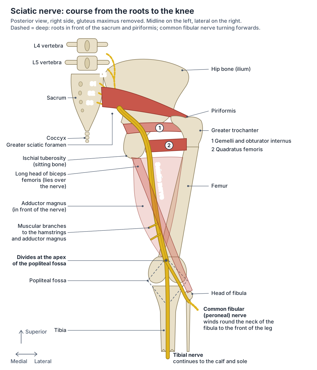
Where is the sciatic nerve located?
The sciatic nerve starts near the base of the spine in the lower back, passes through the pelvis and the buttock, and runs down the back of each thigh into the lower leg. There is one on each side. Even though it passes through the buttock, it does not control the buttock muscles. (Cleveland Clinic: Sciatica.)
In more detail, the nerve leaves the pelvis through the greater sciatic foramen and enters the buttock (the gluteal region) just below the piriformis muscle. It then runs down and outwards and enters the back of the thigh by passing deep to the long head of biceps femoris. At the top (apex) of the popliteal fossa, the hollow behind the knee, it ends by dividing into the tibial and common fibular nerves. (TeachMeAnatomy: The Sciatic Nerve.)
In the buttock the nerve runs down between the ischial tuberosity (the sitting bone) and the greater trochanter at the top of the femur. Textbooks place it about midway between the two. A 2024 dissection study of 62 buttocks from 31 adult Chinese cadavers (24 men and 7 women) found that the answer depends on which point of the tuberosity is used: measured from its inner (medial) edge, the nerve lay at about the midpoint, in agreement with the textbooks; measured from its most prominent point, it lay about a third of the way across, nearer the tuberosity. Its exact position varied between people. (Ma C, Wang X, Li J. Assessment in location of sciatic nerve between the ischial tuberosity and the greater trochanter of the femur. Heliyon. 2024.)
The nerve's path explains a familiar rule. If the buttock is divided into four quadrants, the sciatic nerve passes through the lower inner quadrant, so intramuscular injections into the buttock are given only in the upper outer quadrant. (TeachMeAnatomy: The Sciatic Nerve.)
The layers of the gluteal region, the gluteal vessels and nerves, and the safe injection sites. Buttocks anatomy.
Sciatic nerve roots: L4 to S3
The sciatic nerve is a branch of the sacral plexus. It is formed from the anterior (ventral) rami of five spinal nerves, L4, L5, S1, S2 and S3, and carries fibres from both the anterior and the posterior divisions of these rami. The five roots converge on the back wall of the pelvis to form a single large nerve. (Kenhub: Sciatic nerve.)
The sacral plexus lies on the posterior wall of the pelvis, in front of piriformis. It is formed by the anterior rami of S1 to S4, joined by the 4th and 5th lumbar roots, which combine as the lumbosacral trunk and descend into the pelvis to meet the sacral roots. (TeachMeAnatomy: The Sacral Plexus.)
Radiopaedia gives the same origin, from the anterior rami of the fourth lumbar to the third sacral spinal nerves, and explains that the tibial part of the nerve is made of the anterior divisions of these rami. It describes the sciatic nerve as the largest and widest nerve in the body, typically about 2 cm wide. (Radiopaedia: Sciatic nerve.)
The common fibular (peroneal) part, by contrast, contains mainly fibres from the posterior divisions of the sacral plexus, and its root value is L4 to S2. (Kenhub: Common fibular (peroneal) nerve.)
So the sciatic nerve is really two nerves bundled together in one connective tissue sheath: the tibial nerve and the common fibular nerve. TeachMeAnatomy describes the bundle as a thick, flat band about 2 cm wide, the largest nerve in the body. (TeachMeAnatomy: The Sciatic Nerve.)
The lumbar and sacral plexuses and every major nerve of the leg, with a skin territory map. Nerves of the lower limb.
The two parts of the sciatic nerve
| Part | Roots | Divisions of the anterior rami | Becomes |
|---|---|---|---|
| Tibial part | L4 to S3 | Anterior divisions | Tibial nerve: back of the leg and sole of the foot |
| Common fibular (peroneal) part | L4 to S2 | Mainly posterior divisions | Common fibular nerve: front and side of the leg and top of the foot |
Roots from TeachMeAnatomy and Kenhub (tibial and common fibular nerves); divisions from Radiopaedia (tibial) and Kenhub (common fibular). The whole sciatic nerve carries L4 to S3.
Greater sciatic foramen: where the sciatic nerve leaves the pelvis
The greater sciatic foramen is the large opening in the back of the pelvis through which structures pass from the pelvis into the gluteal region. It is bordered by the sacrotuberous ligament behind and medially, the greater sciatic notch of the ilium in front and laterally, and the sacrospinous ligament and ischial spine below. (TeachMeAnatomy: The Greater and Lesser Sciatic Foramina.)
OpenStax describes the same opening as formed by the greater sciatic notch of the hip bone, the sacrum and the sacrospinous ligament, with the smaller lesser sciatic foramen below it. (OpenStax Anatomy and Physiology 2e, 8.3: The Pelvic Girdle and Pelvis.)
Piriformis passes out through the foramen and divides it into two parts. Above piriformis (the suprapiriform foramen) pass the superior gluteal artery, vein and nerve. Below piriformis (the infrapiriform foramen) pass the sciatic nerve, the pudendal nerve, the inferior gluteal artery, vein and nerve, the posterior femoral cutaneous nerve, the nerve to obturator internus and the nerve to quadratus femoris. (TeachMeAnatomy: The Greater and Lesser Sciatic Foramina.)
Kenhub lists the same companions for the sciatic nerve as it passes below piriformis: the posterior femoral cutaneous nerve, the pudendal nerve, the internal pudendal artery and vein, and the inferior gluteal nerve, artery and vein. (Kenhub: Sciatic nerve.)
What passes through the greater sciatic foramen
| Position | Structure | Main area served |
|---|---|---|
| Above piriformis | Superior gluteal nerve (L4 to S1) | Gluteus medius, gluteus minimus and tensor fasciae latae |
| Above piriformis | Superior gluteal artery and vein | Run with the superior gluteal nerve |
| Below piriformis | Sciatic nerve (L4 to S3) | Back of the thigh, the whole leg and foot |
| Below piriformis | Inferior gluteal nerve (L5 to S2) | Gluteus maximus |
| Below piriformis | Inferior gluteal artery and vein | Gluteal muscles, head of the femur, the sciatic nerve and the upper back of the thigh |
| Below piriformis | Posterior femoral cutaneous nerve (S1 to S3) | Skin of the buttock, the back of the thigh, the upper calf and the perineum |
| Below piriformis | Pudendal nerve (S2 to S4) with the internal pudendal artery and vein | Perineum; they leave through the greater and re-enter through the lesser sciatic foramen |
| Below piriformis | Nerve to obturator internus; nerve to quadratus femoris | Obturator internus and superior gemellus; quadratus femoris and inferior gemellus |
Contents from TeachMeAnatomy (sciatic foramina, sacral plexus, inferior gluteal artery) and Kenhub (sciatic nerve, sacral plexus); skin supply of the posterior femoral cutaneous nerve also from Radiopaedia.
Sciatic nerve course: buttock, thigh and back of the knee
After leaving the pelvis below piriformis, the nerve descends in an inferolateral direction through the gluteal region. It crosses the back of four small hip rotators in turn: superior gemellus, obturator internus, inferior gemellus and quadratus femoris. It does not supply any of the muscles of the gluteal region. (TeachMeAnatomy: The Sciatic Nerve.)
Radiopaedia describes the same layers: in its descent the nerve lies deep to gluteus maximus and behind obturator internus, the gemelli and quadratus femoris. Lower down it lies behind adductor magnus and is crossed from behind by the long head of biceps femoris. (Radiopaedia: Sciatic nerve.)
In the thigh the nerve runs between the long head of biceps femoris and adductor magnus, lateral to semitendinosus and semimembranosus. On the way it gives small muscular branches to the muscles of the back of the thigh and to the ischial (hamstring) part of adductor magnus. (Kenhub: Sciatic nerve.)
In the buttock the nerve lies deep to gluteus maximus and to the inferior gluteal artery, and it receives small vessels (vasa nervorum) from that artery. (Kenhub: Sacral plexus.)
TeachMeAnatomy likewise lists the sciatic nerve among the structures supplied by the inferior gluteal artery, together with the gluteal muscles, the head of the femur and the skin and muscles of the upper back of the thigh. (TeachMeAnatomy: Inferior Gluteal Artery.)
Behind the knee, the tendons of the hamstring muscles (biceps femoris, semitendinosus and semimembranosus) form the upper borders of the popliteal fossa, the diamond-shaped space at the back of the knee. (OpenStax Anatomy and Physiology 2e, 11.6: Appendicular Muscles of the Pelvic Girdle and Lower Limbs.)
The hamstrings and adductor magnus that the sciatic nerve runs between and supplies. Thigh muscles.
Sciatic nerve and piriformis: the Beaton and Anson types
The relation between the sciatic nerve and piriformis was classified by Beaton and Anson in 1937 into six types. In type A, the usual pattern, the undivided nerve passes below piriformis. In type B the common fibular (peroneal) division passes through the muscle and the tibial division below it; in type C the common fibular division passes above and the tibial division below; in type D the undivided nerve passes through the muscle; in type E the common fibular division passes above and the tibial division through it; and in type F the undivided nerve passes above piriformis. (Poutoglidou F et al. Sciatic nerve variants and the piriformis muscle: a systematic review and meta-analysis. Cureus. 2020.)
A 2022 cadaver study gives the same six types in Beaton and Anson's original wording, in which types D and E describe a piriformis split into two heads: in type D the undivided nerve passes between the heads, and in type E one division passes between the heads and the other above. (Reynoso JP et al. Anatomical variations of the sciatic nerve exit from the pelvis and its relationship with the piriformis muscle. Neurol Int. 2022.)
How common are these patterns? A 2020 meta-analysis pooled 44 cadaver studies with 8,257 specimens. The typical type A pattern was found in 90%, type B in 8%, type C in 2% and type D in 1%, and some variant in 13%. Beaton and Anson described types E and F as theoretical, and they were too rarely reported to be pooled, but single cases of both have since been found in dissections; the 2022 study below found one type E pattern in 40 limbs. Variants were commonest in East Asian populations, with a pooled prevalence of 31%. (Poutoglidou F et al. Sciatic nerve variants and the piriformis muscle: a systematic review and meta-analysis. Cureus. 2020.)
An earlier meta-analysis of 18 studies and 6,062 cadavers found a piriformis or sciatic nerve variant in 16.9%. Variants were found about as often (16.2%) in surgical series of people operated on for piriformis syndrome, which suggests they may matter less in causing the syndrome than had been thought. (Smoll NR. Variations of the piriformis and sciatic nerve with clinical consequence: a review. Clin Anat. 2010.)
Single studies agree that the typical pattern dominates. In the 2022 study of 40 sciatic nerves, 37 (92.5%) left the pelvis below piriformis. The same paper cites a series of 294 limbs in which 93.6% had the typical pattern and the commonest variant, in 4.1%, was a common fibular division passing through piriformis with the tibial division below it. (Reynoso JP et al. Anatomical variations of the sciatic nerve exit from the pelvis and its relationship with the piriformis muscle. Neurol Int. 2022.)
The two divisions develop separately in the embryo, which may explain why they can take different routes around the muscle. Knowing the variants matters in hip replacement through a posterior approach, in sciatic nerve blocks and in injections guided into piriformis. (Poutoglidou F et al. Sciatic nerve variants and the piriformis muscle: a systematic review and meta-analysis. Cureus. 2020.)
Piriformis, the gemelli, obturator internus, quadratus femoris and the gluteal muscles above them. Hip muscles.
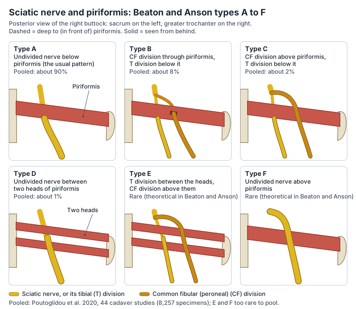
Beaton and Anson types: how the sciatic nerve passes piriformis
| Type | Route | Pooled prevalence (2020 meta-analysis) |
|---|---|---|
| A | Undivided nerve below piriformis (typical) | 90% |
| B | Common fibular division through piriformis, tibial division below | 8% |
| C | Common fibular division above piriformis, tibial division below | 2% |
| D | Undivided nerve through piriformis (between two heads) | 1% |
| E | Common fibular division above, tibial division through (between two heads) | Rare; not pooled |
| F | Undivided nerve above piriformis | Rare; not pooled |
Types from Beaton and Anson (1937) as given by Poutoglidou et al. 2020 and Reynoso et al. 2022; type E division routes also in Radiopaedia. Pooled prevalence: Poutoglidou et al. 2020, 44 studies, 8,257 specimens.
Sciatic nerve branches
The sciatic nerve gives three kinds of branches. Articular branches go to the hip and knee joints. Muscular branches go to biceps femoris, semitendinosus, semimembranosus and the hamstring (ischiocondylar) part of adductor magnus. Its two terminal branches are the tibial and common fibular nerves. (Radiopaedia: Sciatic nerve.)
Spine-health describes the articular branches more precisely: they supply the back of the hip joint and the back and side of the knee joint. (Spine-health: Sciatic Nerve Anatomy.)
The muscular branches come from the two parts of the nerve. The tibial part supplies the long head of biceps femoris, semitendinosus and semimembranosus. The short head of biceps femoris, which arises from the femur rather than the ischial tuberosity, is supplied by the common fibular part. (TeachMeAnatomy: Muscles of the Posterior Thigh (Hamstrings).)
The tibial and common fibular parts usually separate at the apex of the popliteal fossa. In about 12% of people, though, they are already separate as they leave the pelvis. (TeachMeAnatomy: The Sciatic Nerve.)
A 2026 meta-analysis of 20 cadaver studies (1,684 lower limbs) found that about 31% of sciatic nerves divided somewhere other than the usual site, roughly one in three. Division inside the pelvis was found in about 11%, close to the TeachMeAnatomy figure. (Liyew WA, Abebe S. Anatomical variation of the sciatic nerve division in the cadaver studies: a systematic review and meta-analysis. Anat Cell Biol. 2026.)
Radiopaedia explains why: the two nerves are only loosely combined as the sciatic nerve, so they can separate at any point between the sacral plexus and the lower third of the thigh. Most often the division lies above the knee, close to it. (Radiopaedia: Sciatic nerve.)
The tibial nerve (L4 to S3) arises at the apex of the popliteal fossa and continues down the back of the leg. It passes behind and below the medial malleolus through the tarsal tunnel, gives medial calcaneal branches to the skin of the heel, and divides into the medial and lateral plantar nerves of the sole. (TeachMeAnatomy: The Tibial Nerve.)
Kenhub gives the same root value, L4 to S3, and describes the tibial nerve as the larger of the two terminal branches. It usually arises in the lower third of the back of the thigh, close to the apex of the popliteal fossa. (Kenhub: Tibial nerve.)
The common fibular (peroneal) nerve (L4 to S2) follows the medial border of biceps femoris down and outwards over the lateral head of gastrocnemius. It wraps around the neck of the fibula and divides there into the superficial and deep fibular nerves. (TeachMeAnatomy: The Common Fibular Nerve.)
The popliteal fossa, where the sciatic nerve divides, and the hamstring tendons around it. Knee joint.
Sciatic nerve branches
| Branch | Where it arises | Supplies |
|---|---|---|
| Articular branches | Along the course of the nerve | Back of the hip joint; back and side of the knee joint |
| Muscular branches, tibial part | Back of the thigh | Long head of biceps femoris, semitendinosus, semimembranosus, hamstring part of adductor magnus |
| Muscular branch, common fibular part | Back of the thigh | Short head of biceps femoris |
| Tibial nerve (L4 to S3) | Usually the apex of the popliteal fossa | Muscles of the back of the leg and most of the sole; skin of the heel and sole, and of the posterolateral leg and lateral foot with the common fibular nerve (sural nerve) |
| Common fibular (peroneal) nerve (L4 to S2) | Usually the apex of the popliteal fossa | Muscles of the front and side of the leg and extensor digitorum brevis; skin of the lateral leg and the top of the foot |
Sources: Radiopaedia and Spine-health (articular branches); TeachMeAnatomy (hamstrings, tibial and common fibular nerves, sacral plexus); Kenhub (tibial, common fibular and sural nerves).
What does the sciatic nerve supply? Muscles and skin
Although the sciatic nerve passes through the buttock, it supplies no gluteal muscle. It directly supplies the muscles of the back of the thigh and the hamstring part of adductor magnus, whose other part is supplied by the obturator nerve, and through its two terminal branches it supplies every muscle of the leg and foot. (TeachMeAnatomy: The Sciatic Nerve.)
Through the tibial nerve it supplies the muscles of the back of the leg: gastrocnemius, soleus, plantaris, popliteus, flexor hallucis longus, flexor digitorum longus and tibialis posterior, and then most of the muscles of the sole. Through the common fibular nerve it supplies the front of the leg (tibialis anterior, extensor hallucis longus, extensor digitorum longus and fibularis (peroneus) tertius) and the side of the leg (fibularis longus and brevis). (Kenhub: Sciatic nerve.)
TeachMeAnatomy summarises the two parts the same way. The tibial part supplies the back of the thigh except the short head of biceps femoris, the hamstring part of adductor magnus, all the muscles of the back of the leg and the sole. The common fibular part supplies the short head of biceps femoris, all the muscles of the front and side of the leg, and extensor digitorum brevis on the top of the foot. (TeachMeAnatomy: The Sacral Plexus.)
The sciatic nerve has no direct skin branches. Through its terminal branches it supplies the skin of the lateral leg, the heel and both the top and the sole of the foot: the tibial nerve covers the posterolateral leg, the lateral foot and the sole, and the common fibular nerve the lateral leg and the top (dorsum) of the foot. (TeachMeAnatomy: The Sciatic Nerve.)
In more detail, the superficial fibular nerve supplies the skin of the anterolateral leg and most of the top of the foot, the deep fibular nerve a small area between the big and second toes, and the lateral sural cutaneous nerve the skin on the outer side of the leg just below the knee. (Kenhub: Common fibular (peroneal) nerve.)
The sural nerve, formed from branches of both the tibial and the common fibular nerves, is purely sensory. It supplies the posterolateral skin of the lower third of the leg and the outer side of the foot, heel and ankle. (Kenhub: Sural nerve.)
One gap often surprises students: the skin on the back of the thigh is not supplied by the sciatic nerve. It belongs to the posterior femoral cutaneous nerve, a separate branch of the sacral plexus that leaves the pelvis below piriformis with the sciatic nerve and supplies the skin of the buttock, the back of the thigh and the upper back of the calf. (Radiopaedia: Posterior femoral cutaneous nerve.)
What the sciatic nerve supplies, region by region
| Region | Muscles | Skin | Through |
|---|---|---|---|
| Buttock | None (it only passes through) | None (posterior femoral cutaneous and other nerves) | Not applicable |
| Back of the thigh | Biceps femoris (both heads), semitendinosus, semimembranosus, hamstring part of adductor magnus | None (posterior femoral cutaneous nerve) | Sciatic nerve directly |
| Back of the leg (calf) | Gastrocnemius, soleus, plantaris, popliteus, tibialis posterior, flexor digitorum longus, flexor hallucis longus | Posterolateral lower leg (sural nerve) | Tibial nerve |
| Front of the leg | Tibialis anterior, extensor hallucis longus, extensor digitorum longus, fibularis tertius | Anterolateral leg (superficial fibular nerve) | Common fibular nerve (deep fibular branch for the muscles) |
| Side of the leg | Fibularis (peroneus) longus and brevis | Outer side of the leg just below the knee (lateral sural cutaneous nerve) | Common fibular nerve (superficial fibular branch for the muscles) |
| Top of the foot | Extensor digitorum brevis | Most of the dorsum (superficial fibular nerve); skin between the big and second toes (deep fibular nerve); outer edge (sural nerve) | Common fibular nerve, with the sural nerve |
| Sole of the foot | Most intrinsic muscles of the foot | Sole (medial and lateral plantar nerves); heel (medial calcaneal branches) | Tibial nerve |
Compiled from TeachMeAnatomy (sciatic, tibial and common fibular nerves, sacral plexus), Kenhub (sciatic, tibial, common fibular and sural nerves) and Radiopaedia (posterior femoral cutaneous nerve).
Where is the sciatic nerve on a woman? Women, men, left and right
Anatomy sources describe the same course for everyone: the sciatic nerve starts near the base of the spine, passes through the pelvis and buttock and runs down the back of each thigh into the lower leg, with one nerve on each side. (Cleveland Clinic: Sciatica.)
What differs is the pelvis around it. The female pelvis is broader, and the greater sciatic notch, which forms the front of the greater sciatic foramen, is broader in females and narrower and deeper in males. (OpenStax Anatomy and Physiology 2e, 8.3: The Pelvic Girdle and Pelvis.)
A University of Wisconsin anatomy lab gives the same contrast, a wider greater sciatic notch in female and a narrower one in male skeletons, and stresses that these are average differences with plenty of overlap between individuals. (University of Wisconsin-Madison, Hominin virtual lab: Pelvis sexual dimorphism.)
Variants around piriformis were slightly more common in female specimens in the 2020 meta-analysis (18% against 11%), but this figure came from only three studies with 290 specimens, and the difference was not statistically significant. (Poutoglidou F et al. Sciatic nerve variants and the piriformis muscle: a systematic review and meta-analysis. Cureus. 2020.)
The 2022 study of 40 nerves found the same trend, with variants in 10% of female and 5% of male limbs, and also judged the numbers too small to be statistically significant. (Reynoso JP et al. Anatomical variations of the sciatic nerve exit from the pelvis and its relationship with the piriformis muscle. Neurol Int. 2022.)
Sciatica is felt on the same side as the irritated nerve or root: irritation of the left sciatic nerve or its roots causes pain in the left buttock and leg, and irritation of the right nerve or its roots pain on the right, in women and men alike. Kenhub describes the typical pain as felt in the ipsilateral (same-side) buttock, radiating down the lower limb. (Kenhub: Sciatic nerve.)
The two sides are also alike in their variants. In the 2020 meta-analysis, piriformis variants were about equally common on the left (23%) and the right (22%) (nine studies, 2,572 specimens), and no significant differences were found by sex or side. (Poutoglidou F et al. Sciatic nerve variants and the piriformis muscle: a systematic review and meta-analysis. Cureus. 2020.)
One rare cause of sciatic pain occurs only in women: endometriosis growing into the sciatic nerve or the sacral plexus. A 2026 review found that nerve-invasive endometriosis mainly involves the sciatic nerve and that the hallmark clue is cyclical (catamenial) pain, pins and needles or weakness that worsens around menstruation. It advises that this cause be considered in any woman with cyclical sciatic, buttock or pudendal pain, because timely diagnosis can prevent permanent nerve damage. (de Leeuw R et al. Aetiology of pelvic neuropathies in women: a narrative review. Facts Views Vis Obgyn. 2026.)
Isolated endometriosis of the sciatic nerve is considered extremely rare, and its symptoms overlap with those of ordinary sciatica, which makes it difficult to recognise. (Zamurovic M et al. Isolated deep infiltrating endometriosis of the sciatic nerve: a case report and overview of the literature. Medicina. 2023.)
The hip bones, the sciatic notches and the differences between the male and female pelvis. Pelvis.
Sciatica vs sciatic nerve: what is sciatic nerve pain?
The sciatic nerve is a structure; sciatica is a symptom. OpenStax describes sciatica as the result of compression or irritation of the sciatic nerve or of any of the spinal nerves that give rise to it. (OpenStax Anatomy and Physiology 2e, 13.4: The Peripheral Nervous System.)
The NHS describes sciatica as what happens when the sciatic nerve, which runs from the lower back to the feet, is irritated or compressed. It names a slipped (herniated) disc as the most common cause, with spinal stenosis (narrowing of the space the nerves pass through), spondylolisthesis (a vertebra slipping out of position) and back injuries as other causes. (NHS: Sciatica.)
In most people the problem therefore lies at the nerve roots in the lower spine rather than in the nerve in the buttock or thigh. A herniated disc pressing on a nerve root is the usual cause, and the medical literature also calls sciatica lumbosacral radicular syndrome or nerve root pain. (Koes BW, van Tulder MW, Peul WC. Diagnosis and treatment of sciatica. BMJ. 2007.)
Cleveland Clinic separates true sciatica, in which an injury or condition directly affects the sciatic nerve, from sciatica-like conditions, which feel the same but come from nearby nerves or from the group of nerves that form the sciatic nerve. (Cleveland Clinic: Sciatica.)
Two related terms are worth keeping apart. Radicular pain is shooting or burning pain that travels down the limb because a single nerve root or its ganglion is irritated; radiculopathy is a measurable loss of sensation or strength because conduction along the root is impaired. The two often occur together, and lumbar radicular pain is what is commonly called sciatica. (Kenhub: Dermatomes.)
The dermatome map from C2 to S5, and why radicular pain follows dermatomes only roughly. Dermatomes.
Where is sciatic nerve pain felt, and why?
Sciatica usually affects the buttock and the back of one leg, often down into the foot and toes. It can feel like a sharp, burning pain down the back of the leg, with pins and needles, numbness or weakness, and it may be worse when moving, sneezing or coughing. There may be back pain too, but it is usually not as bad as the leg pain. (NHS: Sciatica.)
Cleveland Clinic describes the pain as burning or like an electric shock shooting down one leg, felt in the lower back, buttock or leg. Sciatica usually affects one leg at a time and only rarely both. (Cleveland Clinic: Sciatica.)
A 2024 review by the World Federation of Neurosurgical Societies describes the same pattern for a lumbar disc herniation: pain often starts in the lower back, can spread to the buttocks and then radiates into the leg, usually on one side only. Most of these herniations in adults aged 25 to 55 are at the L4-L5 or L5-S1 level. (Pojskic M et al. Lumbar disc herniation: epidemiology, clinical and radiologic diagnosis. WFNS spine committee recommendations. World Neurosurg X. 2024.)
OpenStax also names the L4/L5 and L5/S1 discs as the most common sites of disc herniation and links them with sciatica, a widespread pain that radiates from the lower back down the thigh and into the leg. (OpenStax Anatomy and Physiology 2e, 7.3: The Vertebral Column.)
The pain is felt in the leg, not just where the root is pinched, because sciatica is radiating pain that follows a dermatomal pattern: the strip of skin supplied by the irritated nerve root. (Koes BW, van Tulder MW, Peul WC. Diagnosis and treatment of sciatica. BMJ. 2007.)
Which root is irritated depends on where the disc bulges. A paracentral herniation, backwards and to one side, presses on the root that crosses the disc on its way down to the next foramen, while a far lateral herniation presses on the root leaving at that level. A paracentral herniation at L4-L5 therefore causes an L5 radiculopathy, and a far lateral herniation at the same level an L4 radiculopathy. (Amin RM, Andrade NS, Neuman BJ. Lumbar disc herniation. Curr Rev Musculoskelet Med. 2017.)
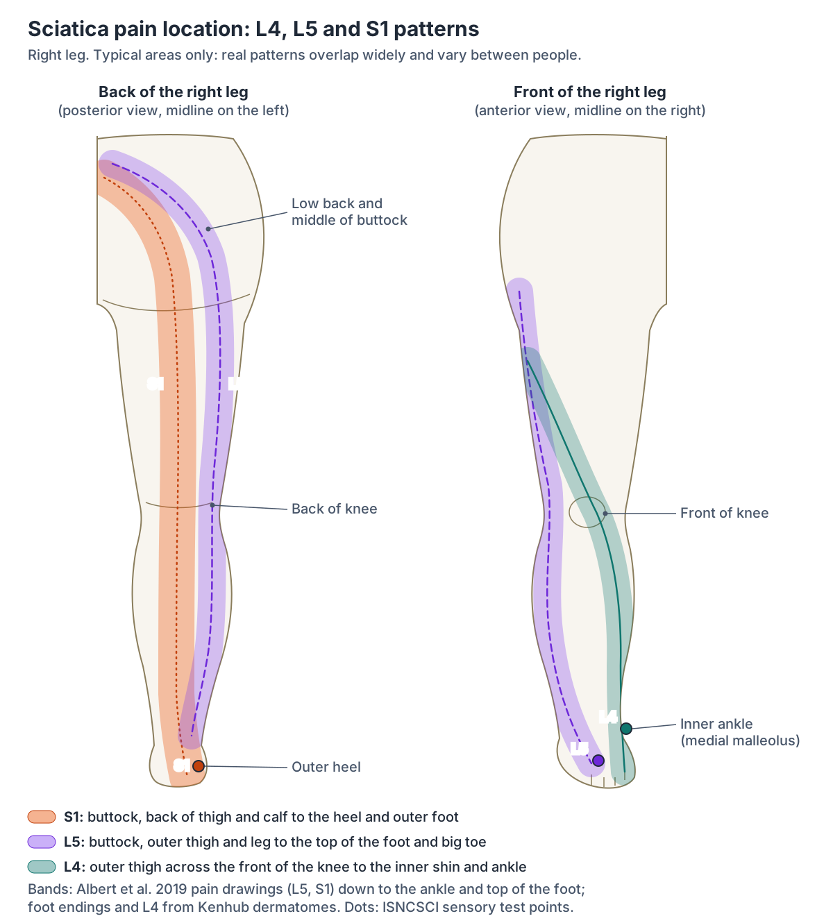
In a Danish study of 93 people with an MRI-confirmed single-level disc herniation, L5 root pain ran from the centre of the low back diagonally across the middle of the buttock and down the outer back of the thigh and calf to the ankle, and also along the outer front of the thigh and calf to the top of the foot. S1 root pain took the same path across the buttock and then ran down the back of the thigh and calf to the ankle. (Albert HB et al. Where do patients with MRI-confirmed single-level radiculopathy experience pain? Chiropr Man Therap. 2019.)
Dermatome maps fill in the foot. L4 covers the knee, the inner leg, the inner ankle and the inner side of the big toe; L5 the outer leg, the top and sole of the foot and the middle toes; and S1 the outer ankle, the outer edge of the foot, the heel and the little toe. (Kenhub: Dermatomes.)
Clinicians test sensation for these roots at standard points: the inner ankle (medial malleolus) for L4, the top of the foot at the third metatarsophalangeal joint for L5, and the outer heel for S1. (Kirshblum SC et al. International standards for neurological classification of spinal cord injury (revised 2011). J Spinal Cord Med. 2011.)
These patterns are only a guide. In the Danish study, pain patterns overlapped published dermatomes by only about 50 to 80%, and clinicians who tried to name the root from pain drawings alone were right about as often as chance (54.4 to 54.8%). (Albert HB et al. Where do patients with MRI-confirmed single-level radiculopathy experience pain? Chiropr Man Therap. 2019.)
Get urgent help (in the UK, go to A&E or call 999) if sciatica affects both legs, if both legs become severely weak or numb, if there is numbness around the genitals or anus, or if control of the bladder or bowel changes: these can be signs of a serious back problem that needs hospital treatment. See a GP if the pain is not improving after a few weeks, is getting worse or is stopping normal activities. (NHS: Sciatica.)
The lumbar vertebrae, intervertebral discs and foramina where the L4 to S3 roots leave the spine. Spine anatomy.
Sciatic nerve pain in the buttocks: piriformis syndrome and deep gluteal syndrome
Buttock pain is part of the usual sciatica picture. Kenhub describes sciatica as pain in the buttock on the affected side that radiates down the lower limb, usually caused by a disc herniation in the lumbosacral spine. (Kenhub: Sciatic nerve.)
The buttock can hurt even though the sciatic nerve supplies nothing there, because pain may be referred to the buttock when the nerve is impaired. (Spine-health: Sciatic Nerve Anatomy.)
Less often the nerve is irritated in the buttock itself. Piriformis syndrome is compression of the sciatic nerve by the piriformis muscle, with radiating pain, numbness, weakness and tenderness in the buttock; the pain may be worse when the hip is turned inwards. X-ray and MRI are usually normal but help to rule out problems in the spine. (TeachMeAnatomy: The Sciatic Nerve.)
Piriformis syndrome has been described for over 70 years but remains controversial. A 2010 systematic review of 55 studies, mostly case series, found the commonest reported features to be buttock pain, tenderness over the greater sciatic notch, pain made worse by sitting and pain increased by manoeuvres that tighten piriformis. (Hopayian K et al. The clinical features of the piriformis syndrome: a systematic review. Eur Spine J. 2010.)
Radiopaedia also calls the condition uncommon and controversial, as are its proposed causes, and notes that the nerve and muscle variants blamed for it are seen at a similar rate in the general population. MRI is used mainly to exclude other causes of sciatica. (Radiopaedia: Piriformis syndrome.)
A 2026 review of pelvic nerve problems adds that the label piriformis syndrome is applied inconsistently and lacks agreed diagnostic criteria. (de Leeuw R et al. Aetiology of pelvic neuropathies in women: a narrative review. Facts Views Vis Obgyn. 2026.)
Because the nerve can be trapped at several places in the buttock, many specialists now prefer the broader term deep gluteal syndrome: buttock pain from entrapment of the sciatic nerve outside the pelvis that does not come from a disc. Besides piriformis, the structures involved can include fibrous bands containing blood vessels, the gluteal and hamstring muscles, the gemelli and obturator internus, abnormal blood vessels and space-occupying lesions. (Martin HD, Reddy M, Gómez-Hoyos J. Deep gluteal syndrome. J Hip Preserv Surg. 2015.)
A 2023 review reaches a similar conclusion: it has not been established that piriformis alone entraps the nerve, and pain in the buttock and hamstring region can also come from other nerves, such as the posterior cutaneous nerve of the thigh. (Sharma S et al. Looking beyond piriformis syndrome: is it really the piriformis? Hip Pelvis. 2023.)
Because many different problems cause buttock and leg pain, pain that is not improving after a few weeks or is getting worse should be checked by a GP, and the warning signs listed in the section above need urgent care. (NHS: Sciatica.)
The hip rotator the sciatic nerve crosses, and ischiofemoral impingement, which can mimic sciatica. Quadratus femoris.
Sciatic nerve injury: hip, buttock and injection risks
The nerve itself can be injured by trauma to the buttock, hip fractures, vasculitis, inflammation of muscle, piriformis syndrome, misplaced injections into the buttock and total hip replacement. Complete sciatic nerve palsy is rare. (Radiopaedia: Sciatic nerve.)
Below the knee, the common fibular nerve is the most exposed branch. It lies close to the neck of the fibula and is relatively unprotected, so a fracture there or swelling in the lateral compartment of the leg can damage it, causing foot drop. (Kenhub: Common fibular (peroneal) nerve.)
Damage to the common fibular nerve removes the ability to dorsiflex the foot at the ankle, so the foot hangs permanently plantarflexed (foot drop), and sensation can be lost over the top of the foot and the outer side of the leg. (TeachMeAnatomy: The Common Fibular Nerve.)
The joint the sciatic nerve passes behind, and why a posterior hip dislocation can injure it. Hip joint.
Finding the sciatic nerve's path in the 3D model
The BodyParts3D model used on this site has no nerves of the trunk or limbs, so the sciatic nerve itself is not in the 3D view. The viewer opens from behind and highlights the structures that outline its path: piriformis, the superior and inferior gemelli, obturator internus, quadratus femoris, the long and short heads of biceps femoris, semitendinosus, semimembranosus and adductor magnus. Gluteus maximus is not highlighted, so the deep layer stays visible.
To trace the nerve, find the lower border of piriformis and follow a line down across the back of superior gemellus, obturator internus, inferior gemellus and quadratus femoris, then down the back of the thigh deep to the long head of biceps femoris to the back of the knee. The course diagram at the top of this page shows the same path with the nerve drawn in. (TeachMeAnatomy: The Sciatic Nerve.)
The bones, muscle compartments and nerves of the whole lower limb. Leg anatomy.
Sciatic nerve in the 3D model
- Adductor magnus
- The largest adductor, with an adductor part and a hamstring part.
- Gemellus inferior
- A small lateral rotator of the hip, below obturator internus.
- Gemellus superior
- A small lateral rotator of the hip, above obturator internus.
- Long head of biceps femoris
- The long, superficial head of biceps femoris, the lateral hamstring.
- Obturator internus
- A lateral rotator of the hip that arises inside the pelvis and leaves through the lesser sciatic foramen.
- Piriformis
- A pear-shaped lateral rotator of the hip that passes through the greater sciatic foramen.
- Quadratus femoris
- A flat quadrangular deep gluteal muscle below the inferior gemellus.
- Semimembranosus
- A hamstring muscle on the posteromedial thigh with a long flat tendon of origin.
- Semitendinosus
- A hamstring muscle with a long cord-like tendon on the posteromedial thigh.
- Short head of biceps femoris
- The head of biceps femoris that does not cross the hip joint.
Frequently asked questions
Where is the sciatic nerve located?
The sciatic nerve runs from the lower back through the pelvis and buttock and down the back of the thigh, one on each side. It leaves the pelvis through the greater sciatic foramen below piriformis, passes deep to gluteus maximus between the sitting bone and the greater trochanter, and divides at the top of the hollow behind the knee into the tibial and common fibular nerves.
What nerve roots form the sciatic nerve?
Five roots form it: L4, L5, S1, S2 and S3. Their anterior rami join the sacral plexus on the back wall of the pelvis, in front of piriformis, with L4 and L5 arriving as the lumbosacral trunk. The tibial part of the nerve carries L4 to S3 and the common fibular part L4 to S2, which is why lower lumbar and upper sacral root problems cause sciatica.
What are the branches of the sciatic nerve?
It gives articular branches to the hip and knee joints and muscular branches to the hamstrings (biceps femoris, semitendinosus and semimembranosus) and the hamstring part of adductor magnus. Its two terminal branches are the tibial nerve, which supplies the calf and sole, and the common fibular (peroneal) nerve, which supplies the front and side of the leg and the top of the foot.
Where is sciatic nerve pain felt?
Usually in the buttock and down the back of one leg, often into the foot and toes, sometimes with lower back pain that is milder than the leg pain. The exact area depends on the irritated root: L5 pain tends to run down the outer leg to the top of the foot, and S1 pain down the back of the calf to the heel and outer foot, although these patterns overlap widely.
What does sciatic nerve pain feel like?
It is often described as a sharp, burning or electric shock-like pain that shoots down one leg, sometimes with pins and needles, numbness or weakness. Moving, sneezing or coughing can make it worse. Pain in both legs, numbness around the genitals or anus, or new problems controlling the bladder or bowel need urgent medical help.
Is the sciatic nerve in the same place in women?
Yes. Anatomy sources describe the same course for everyone, with one nerve on each side, and piriformis variants are about equally common on the left and right. The female pelvis is broader, with a wider greater sciatic notch, but the nerve still leaves the pelvis below piriformis and runs down the back of the thigh. Piriformis variants may be slightly commoner in women, but studies have not shown a significant difference.
Does the sciatic nerve go through the piriformis?
Usually not. A 2020 meta-analysis of 44 cadaver studies found the undivided nerve passing below piriformis in about 90% of specimens. The commonest variant, in about 8%, is an early split in which the common fibular division passes through piriformis and the tibial division below it. These are the Beaton and Anson types, first classified in 1937.
What is the difference between sciatica and the sciatic nerve?
The sciatic nerve is the large nerve that runs from the lower back to the leg. Sciatica is the pain that results when that nerve, or more often one of the L4 to S3 roots that form it, is irritated or compressed, most commonly by a slipped (herniated) disc. So sciatica is a symptom, and the sciatic nerve is the structure along which it is felt.
References
- TeachMeAnatomy. The Sciatic Nerve.
- TeachMeAnatomy. The Sacral Plexus.
- TeachMeAnatomy. The Greater Sciatic Foramen and Lesser Sciatic Foramen.
- TeachMeAnatomy. The Tibial Nerve.
- TeachMeAnatomy. The Common Fibular Nerve.
- TeachMeAnatomy. Muscles of the Posterior Thigh.
- TeachMeAnatomy. Inferior Gluteal Artery.
- Kenhub. Sciatic nerve: origin, course and branches.
- Kenhub. Sacral plexus.
- Kenhub. Tibial nerve.
- Kenhub. Common fibular (peroneal) nerve.
- Kenhub. Sural nerve.
- Kenhub. Dermatomes.
- Radiopaedia.org. Sciatic nerve.
- Radiopaedia.org. Posterior femoral cutaneous nerve.
- Radiopaedia.org. Piriformis syndrome.
- OpenStax. Anatomy and Physiology 2e, §13.4 The Peripheral Nervous System. Rice University, 2022.
- OpenStax. Anatomy and Physiology 2e, §7.3 The Vertebral Column. Rice University, 2022.
- OpenStax. Anatomy and Physiology 2e, §8.3 The Pelvic Girdle and Pelvis. Rice University, 2022.
- OpenStax. Anatomy and Physiology 2e, §11.6 Appendicular Muscles of the Pelvic Girdle and Lower Limbs. Rice University, 2022.
- University of Wisconsin-Madison, Department of Anthropology. Hominin virtual lab: pelvis sexual dimorphism.
- NHS. Sciatica. Page last reviewed 3 December 2024.
- Cleveland Clinic. Sciatica. Last updated 10 February 2026.
- Jagannathan J. Sciatic Nerve Anatomy. Spine-health.
- Poutoglidou F, Piagkou M, Totlis T, Tzika M, Natsis K. Sciatic Nerve Variants and the Piriformis Muscle: A Systematic Review and Meta-Analysis. Cureus. 2020;12(11):e11531.
- Reynoso JP, De Jesus Encarnacion M, Nurmukhametov R, et al. Anatomical Variations of the Sciatic Nerve Exit from the Pelvis and Its Relationship with the Piriformis Muscle: A Cadaveric Study. Neurol Int. 2022;14(4):894-902.
- Smoll NR. Variations of the piriformis and sciatic nerve with clinical consequence: a review. Clin Anat. 2010;23(1):8-17.
- Liyew WA, Abebe S. Anatomical variation of the sciatic nerve division in the cadaver studies: a systematic review and meta-analysis. Anat Cell Biol. 2026;59(2):219-229.
- Ma C, Wang X, Li J. Assessment in location of sciatic nerve between the ischial tuberosity and the greater trochanter of the femur: A cadaveric study. Heliyon. 2024;10(1):e23751.
- Hopayian K, Song F, Riera R, Sambandan S. The clinical features of the piriformis syndrome: a systematic review. Eur Spine J. 2010;19(12):2095-2109.
- Martin HD, Reddy M, Gómez-Hoyos J. Deep gluteal syndrome. J Hip Preserv Surg. 2015;2(2):99-107.
- Sharma S, Kaur H, Verma N, Adhya B. Looking beyond Piriformis Syndrome: Is It Really the Piriformis? Hip Pelvis. 2023;35(1):1-5.
- de Leeuw R, Groenman FA, Hudelist G, Don EE, de Vries R, Huirne JA. Aetiology of pelvic neuropathies in women: a narrative review. Facts Views Vis Obgyn. 2026;18(3):222-234.
- Zamurovic M, Tomic A, Djordjevic K, et al. Isolated Deep Infiltrating Endometriosis of the Sciatic Nerve: A Case Report and Overview of the Literature. Medicina (Kaunas). 2023;59(12):2161.
- Albert HB, Hansen JK, Søgaard H, Kent P. Where do patients with MRI-confirmed single-level radiculopathy experience pain, and what is the clinical interpretability of these pain patterns? Chiropr Man Therap. 2019;27:50.
- Koes BW, van Tulder MW, Peul WC. Diagnosis and treatment of sciatica. BMJ. 2007;334(7607):1313-1317.
- Amin RM, Andrade NS, Neuman BJ. Lumbar Disc Herniation. Curr Rev Musculoskelet Med. 2017;10(4):507-516.
- Pojskic M, Bisson E, Oertel J, Takami T, Zygourakis C, Costa F. Lumbar disc herniation: Epidemiology, clinical and radiologic diagnosis WFNS spine committee recommendations. World Neurosurg X. 2024;22:100279.
- Kirshblum SC, Burns SP, Biering-Sorensen F, et al. International standards for neurological classification of spinal cord injury (revised 2011). J Spinal Cord Med. 2011;34(6):535-546.
- Mitsuhashi N. et al. (2009). BodyParts3D: 3D structure database for anatomical concepts. Nucleic Acids Research 37, D782–D785. Data © The Database Center for Life Science, CC BY 4.0.
About this page
Drafted with AI assistance from the sources listed above, checked against them and reviewed by Dr Shaista Andleeb; last reviewed on 3 November 2026. The editorial policy explains how sources are chosen and how errors are corrected. To report a mistake, use the contact page.
Related anatomy
- Lower limb nervesLower limb nerves explained: lumbar and sacral plexus diagram, femoral, saphenous, sciatic, tibial, common peroneal…
- DermatomesDermatome anatomy with front and back dermatome maps: every level from C2 to S5, key landmarks, dermatomes vs…
- ButtocksButtocks anatomy with a 3D model and labelled diagrams: surface landmarks, gluteal muscles, sciatic nerve, gluteal…
- Hip musclesHip muscles with a labeled glute muscles diagram and 3D model: gluteus maximus, medius and minimus, TFL, piriformis…
- Thigh musclesThigh muscles explained: quadriceps and sartorius at the front, adductor muscles of the inner thigh, hamstrings at the…
- Vertebral columnSpine anatomy with a labeled spine diagram numbering every vertebra, nerve and disc diagrams, a vertebrae chart…
Explore the whole body in the interactive 3D human anatomy model, or browse the A–Z list of human structures.