

Anatomy for artists: what to learn first
Art teachers usually build a figure from the inside out. A typical order is the gesture of the pose first, then the skeleton as a framework (skull, spine, rib cage, pelvis and limb bones), then the major landmarks, then the rib cage and pelvis as two solid forms joined by the spine, and only then the major muscle groups, with attention to where each muscle attaches and how the joints move. (The Artillerist: Anatomy for Concept Artists: What You Actually Need to Learn.)
Bones are the most reliable reference points. Proko's landmarks lesson notes that bones change little between body types, while fleshy points such as the navel and nipples can be used but vary more. (Proko: Landmarks of the Human Body.)
A bony landmark, in the words of the painter Lea Colie Wight, is a spot where the bone comes closest to the surface and so tells you about the structure under the form. (Artists Network: How to Draw Bodies with Landmarks to Anatomy (Lea Colie Wight).)
Every bone of the skeleton, in a rotatable 3D model, is covered in the guide to the Skeletal system.
Human proportions: how many heads tall is a person?
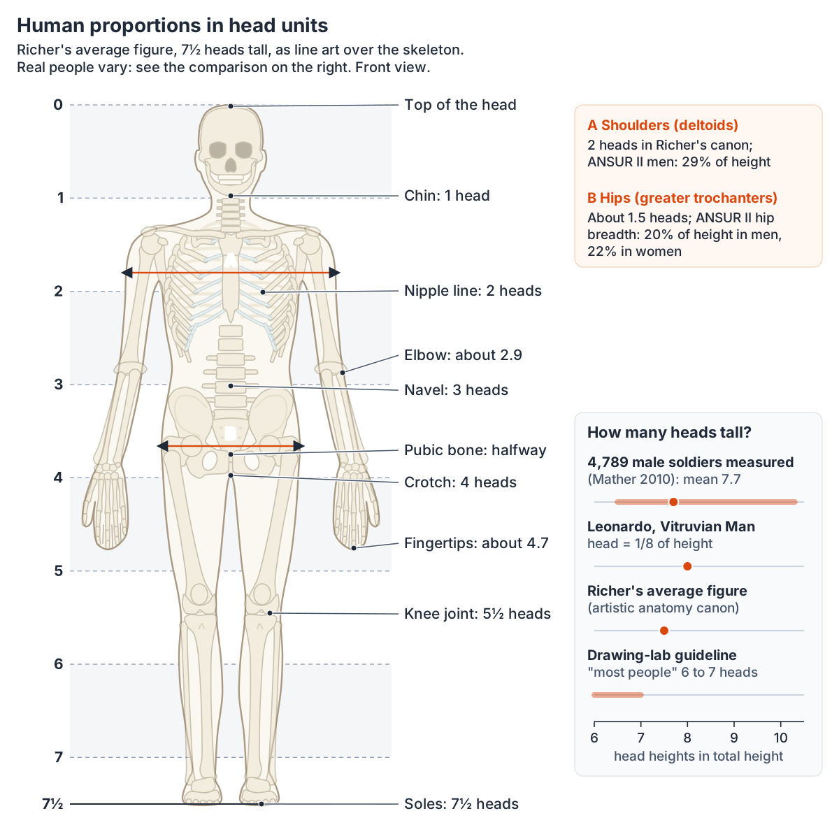
Most figure-drawing systems measure the body in head heights, from the top of the head to the chin. In Paul Richer's artistic anatomy, as taught by Proko, the average person is 7½ heads tall: the head and torso make 4 units and the legs another 4, overlapping by half a unit. The halfway point of the body is at about the pubic bone. (Proko: Human Proportions, Average Figure (lesson notes).)
Richer's landmarks fall on the head lines: one head below the chin are the nipples, the next unit down is the navel (the top of the buttocks at the back), the next is the crotch and the bottom of the buttocks, and the knee joint, at the bottom of the kneecap when the leg is straight, sits halfway down the leg's four units. (Proko: Human Proportions, Average Figure (lesson notes).)
Measurements of real people sit close to these canons but vary a lot. In 4,789 German and NATO male soldiers, head height averaged 0.13 of body height, about 7.7 heads, with a range of 0.097 to 0.153, roughly 6½ to 10 heads. The German soldiers averaged exactly 1/8 (8 heads) and the shorter NATO group 0.132 (about 7.6 heads); taller men tended to have relatively smaller heads. (Mather G. Head-body ratio as a visual cue for stature in people and sculptural art. Perception. 2010;39:1390-1395.)
Sources disagree on the everyday figure. The University of Evansville drawing lab says the average figure is 7 heads high and most people are 6 to 7 heads, but draws its model at 8 heads because that looks more correct in a drawing. (University of Evansville Figure Drawing Lab: Body proportions.)
Leonardo's notes on the Vitruvian Man give the head, from the bottom of the chin to the top of the head, as one eighth of a man's height. (University of Evansville Figure Drawing Lab: Vitruvian proportions.)
Head size changes how tall a figure looks. In an experiment, figures with a smaller head relative to the body were judged taller. Renaissance statues of David by Donatello, Verrocchio and Michelangelo have large heads (head-to-body ratios of 0.14 to 0.16), while statues of Hercules have small ones (0.11 or less), and Michelangelo's David has the head proportion of a man about 165 cm tall. (Mather G. Head-body ratio as a visual cue for stature in people and sculptural art. Perception. 2010;39:1390-1395.)
Labelled whole-body diagrams of the skeleton, muscles and organs, front and back, are collected in the Human body diagram.
Proportions measured in real adults
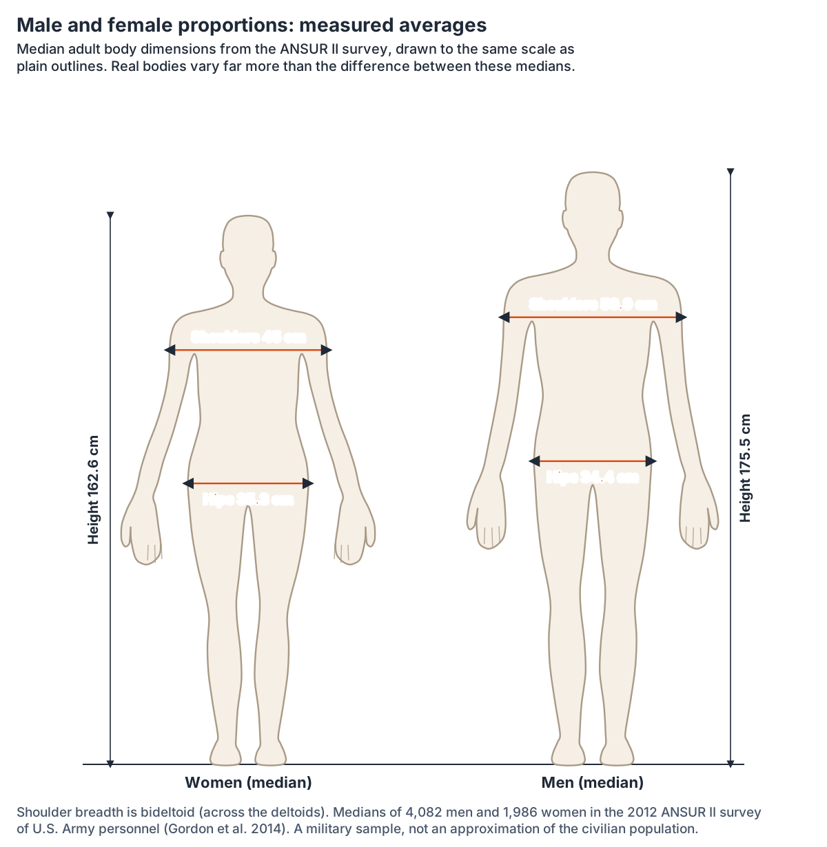
The 2012 ANSUR II survey measured 4,082 men and 1,986 women in the U.S. military. Penn State's OpenLab, which documents the data set, warns that even with reservists included the sample is not an approximation of the U.S. civilian population. (Penn State OpenLab: ANSUR II anthropometric data.)
The median man in ANSUR II is 175.5 cm (69.1 in) tall and the median woman 162.6 cm (64.0 in). His arm span is 181.2 cm and hers 165.7 cm, so the span is slightly longer than height, by about 3 percent in men and 2 percent in women. With the arms hanging, the elbow is at about 62 percent of height above the floor and the fingertips at about 37 percent. (The Ergonomics Center, NC State University: Anthropometric summary tables (ANSUR II, Gordon et al. 2014).)
The civilian figures agree on height: the average U.S. man aged 20 and over is 68.9 in (175 cm) and the average woman 63.5 in (161 cm), measured in 2021 to 2023. (CDC National Center for Health Statistics: FastStats, Body Measurements.)
The canons say much the same about the span. Leonardo wrote that the length of a man's outspread arms is equal to his height. (University of Evansville Figure Drawing Lab: Vitruvian proportions.)
In Richer's figure the width between the fingertips is just a little longer than the height, and is treated as equal in practice. (Proko: Human Proportions, Average Figure (lesson notes).)
Body proportions measured in adults (ANSUR II medians)
| Measurement | Men | Women | Share of height (men / women) |
|---|---|---|---|
| Height (stature) | 175.5 cm | 162.6 cm | 100% / 100% |
| Eye height | 164.0 cm | 151.7 cm | 93.5% / 93.3% |
| Shoulder (acromion) height | 143.9 cm | 133.2 cm | 82.0% / 81.9% |
| Elbow height | 108.3 cm | 100.2 cm | 61.7% / 61.6% |
| Hip height | 89.9 cm | 84.4 cm | 51.2% / 51.9% |
| Fingertip height, arms hanging | 65.4 cm | 61.3 cm | 37.3% / 37.7% |
| Arm span | 181.2 cm | 165.7 cm | 103.3% / 101.9% |
| Shoulder breadth across the deltoids | 50.9 cm | 45.0 cm | 29.0% / 27.7% |
| Shoulder breadth between the acromions | 41.5 cm | 36.5 cm | 23.7% / 22.4% |
| Hip breadth | 34.4 cm | 35.3 cm | 19.6% / 21.7% |
| Elbow to fingertip | 48.0 cm | 43.8 cm | 27.4% / 26.9% |
| Hand length | 19.3 cm | 18.0 cm | 11.0% / 11.1% |
| Foot length | 27.1 cm | 24.4 cm | 15.4% / 15.0% |
Median (50th percentile) values from the 2012 ANSUR II survey of U.S. Army personnel (Gordon et al. 2014), as tabulated by the NC State Ergonomics Center, converted from inches; percentages calculated from the medians. A military sample, not the general population.
Surface anatomy for drawing: bony landmarks on the front
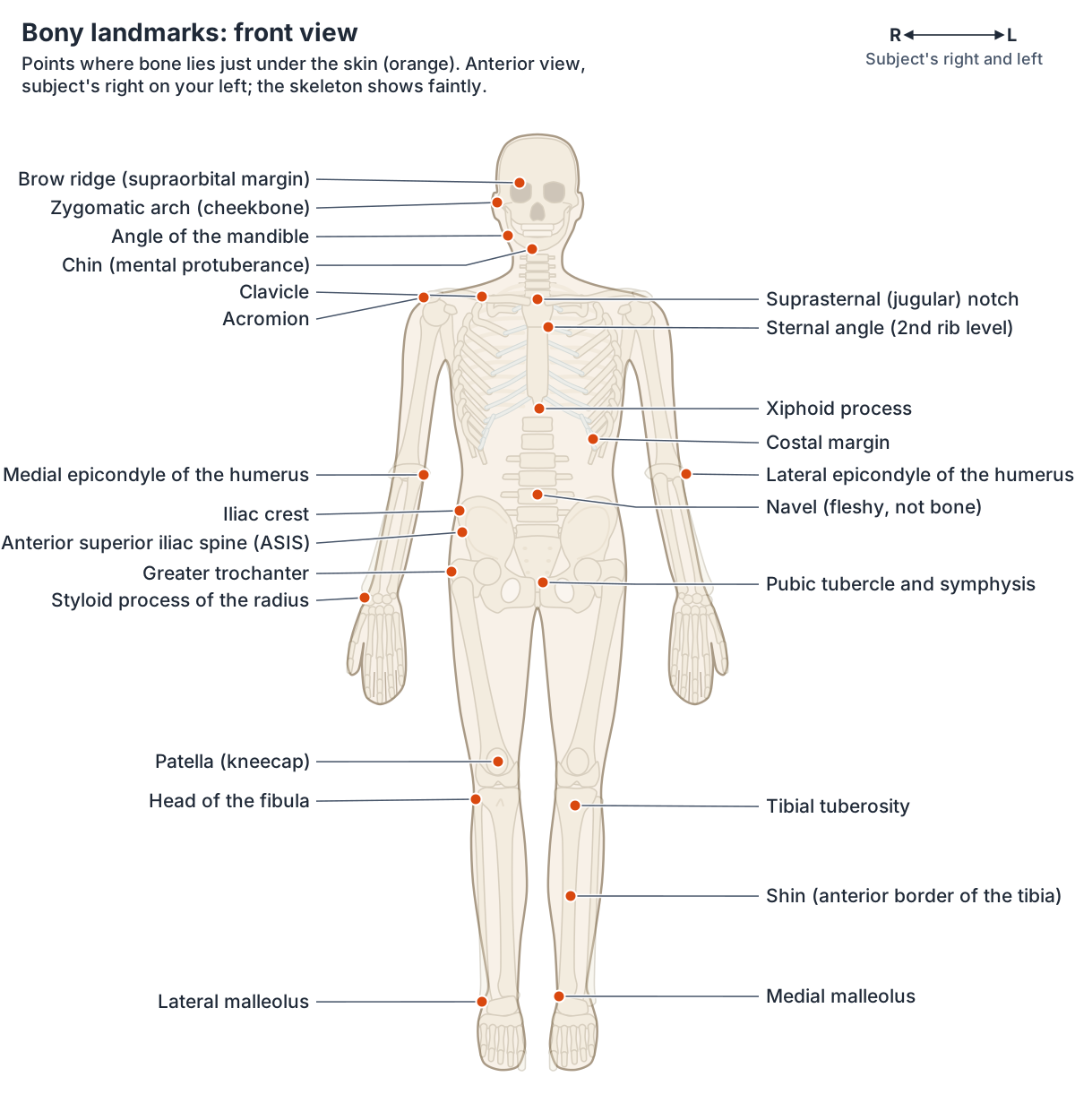
The clavicle (collarbone) is an S-shaped bone on the front of the shoulder that you can feel along its whole length. (OpenStax Anatomy and Physiology 2e, 8.1: The Pectoral Girdle.)
On top of the manubrium of the sternum is the jugular (suprasternal) notch, which you can feel at the base of the neck. Lower down, the sternal angle marks where the second rib joins the sternum; because the first rib is hidden behind the clavicle, the second rib is the highest rib you can feel. (OpenStax Anatomy and Physiology 2e, 7.4: The Thoracic Cage.)
Proko uses the acromion processes, the bony points at the top of the shoulders between the deltoid and the trapezius, to read the angle of the shoulders, and notes that when a shoulder is raised the clavicle rotates from the pit of the neck without changing the angle of the rib cage. (Proko: Landmarks of the Human Body.)
At the elbow, the medial epicondyle of the humerus is the prominent bump on the inner side and the lateral epicondyle the smaller one on the outer side; behind them, the olecranon of the ulna forms the tip of the elbow. At the wrist, the styloid process of the radius points down on the thumb side and the rounded head of the ulna sits on the little-finger side. (OpenStax Anatomy and Physiology 2e, 8.2: Bones of the Upper Limb.)
The iliac crest is the arching upper edge of the hip bone that you feel along the waistline, and its front end, the anterior superior iliac spine (ASIS), can be felt at the front of the hip. (OpenStax Anatomy and Physiology 2e, 8.3: The Pelvic Girdle and Pelvis.)
Proko uses the two ASIS points, the front corners of the iliac crest, to show the angle of the pelvis, and the greater trochanter as the point the gluteal muscles wrap around. (Proko: Landmarks of the Human Body.)
On the outer upper thigh the greater trochanter of the femur can be felt just under the skin. The patella sits in the quadriceps tendon at the front of the knee, the tibial tuberosity below it is where the patellar ligament attaches, and the front ridge of the tibia, the shin, lies immediately under the skin. The ankle shows the medial malleolus of the tibia on the inside and the lateral malleolus of the fibula on the outside. (OpenStax Anatomy and Physiology 2e, 8.4: Bones of the Lower Limb.)
On the abdomen, the linea alba runs from the xiphoid process to the pubic symphysis, and the transverse creases over the rectus abdominis lie over its tendinous intersections. The navel marks about the level of the disc between the third and fourth lumbar vertebrae in people who are not obese. (Texas Tech University Health Sciences Center El Paso: Topographical anatomy of the abdomen.)
Surface regions and landmarks of the front and back of the body are mapped in the guide to Body regions.
Landmarks on the back of the figure
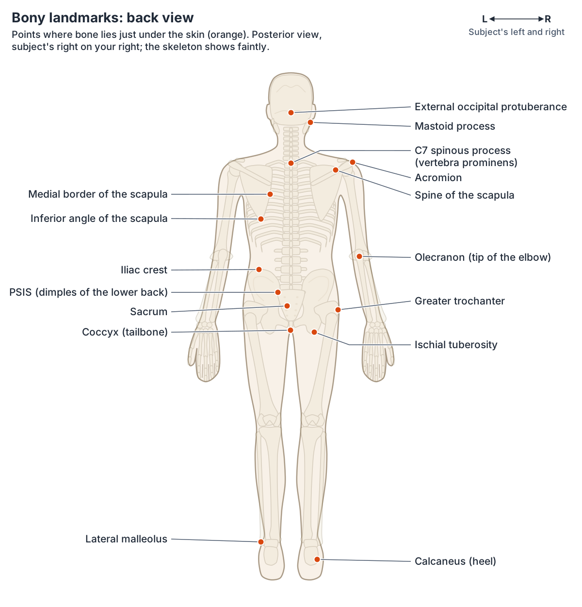
At the base of the neck the spine of the seventh cervical vertebra, the vertebra prominens, is easy to see and feel. Below it the vertebral furrow marks the line of the vertebral spines, with the muscle masses of the erector spinae on either side, and the natal cleft runs between the buttocks. (Texas Tech University Health Sciences Center El Paso: Topographical anatomy of the back.)
The scapula lies on the back of the shoulder. The long, prominent ridge of its spine runs across its upper part, the acromion forms the bony tip of the shoulder, and the lowest point of the scapula is the inferior angle. (OpenStax Anatomy and Physiology 2e, 8.1: The Pectoral Girdle.)
Proko notes that the medial edges of the scapula are not covered by muscle, so they show at the surface, and describes the sacrum as a triangle between the two dimples of the lower back and the tailbone. (Proko: Landmarks of the Human Body.)
The dimples themselves sit over the posterior superior iliac spines, which muscles and ligaments surround without covering. (OpenStax Anatomy and Physiology 2e, 8.3: The Pelvic Girdle and Pelvis.)
On the head, the mastoid process can be felt just behind the earlobe, and the external occipital protuberance is a small bump in the midline at the back of the skull. (OpenStax Anatomy and Physiology 2e, 7.2: The Skull.)
The spine, scapulae and back muscles are described together in Back anatomy.
Bony landmarks for figure drawing
| Landmark | Bone | Where to see or feel it |
|---|---|---|
| Jugular (suprasternal) notch | Manubrium of the sternum | Pit of the neck, between the inner ends of the clavicles |
| Clavicle | Clavicle | Whole length, front of the shoulder |
| Acromion | Scapula | Bony tip of the shoulder |
| Sternal angle | Sternum | Ridge where the second rib joins |
| C7 spinous process | Seventh cervical vertebra | Base of the back of the neck |
| Spine and inferior angle of the scapula | Scapula | Upper back |
| Medial and lateral epicondyles, olecranon | Humerus, ulna | Elbow |
| Styloid process of the radius, head of the ulna | Radius, ulna | Thumb side and little-finger side of the wrist |
| Iliac crest and ASIS | Hip bone | Waistline and front of the hip |
| PSIS | Hip bone | Two dimples of the lower back |
| Greater trochanter | Femur | Outer upper thigh |
| Patella and tibial tuberosity | Patella, tibia | Front of the knee |
| Anterior border of the tibia | Tibia | Shin |
| Medial and lateral malleoli | Tibia, fibula | Inner and outer ankle |
| Mastoid process | Temporal bone | Behind the earlobe |
Compiled from OpenStax Anatomy and Physiology 2e (sections 7.2 to 8.4) and the Texas Tech topographical anatomy tables cited above.
Muscles that shape the torso and neck
The sternocleidomastoid runs from the sternum and clavicle up to the mastoid process behind the ear and divides the side of the neck into anterior and posterior triangles. In the back, the erector spinae group forms most of the muscle mass. (OpenStax Anatomy and Physiology 2e, 11.3: Axial Muscles of the Head, Neck, and Back.)
The deltoid is the thick muscle that creates the rounded line of the shoulder. Pectoralis major is thick and fan-shaped and covers much of the upper chest, the trapezius shrugs the shoulders and draws the shoulder blades together, and the broad, triangular latissimus dorsi covers the lower part of the back. Serratus anterior, on the side of the rib cage, moves the arm from the side of the body to the front. (OpenStax Anatomy and Physiology 2e, 11.5: Muscles of the Pectoral Girdle and Upper Limbs.)
The rectus abdominis is crossed by three tendinous intersections, which make the six-pack segments more prominent with training, and the white linea alba separates the two sides. The external oblique, the outermost abdominal muscle, has fibres that run down and towards the midline. (OpenStax Anatomy and Physiology 2e, 11.4: Axial Muscles of the Abdominal Wall and Thorax.)
Every modelled muscle and the main muscle groups are covered in the guide to the Muscular system.
Muscles that shape the arms and legs
In the arm, the two-headed biceps brachii flexes the forearm and the brachialis lies deep to it; on the back, the triceps brachii extends the forearm. The brachioradialis can flex the forearm quickly or help lift a load slowly. In the palm, the thenar muscles make the rounded base of the thumb and the hypothenar muscles the rounded edge below the little finger. (OpenStax Anatomy and Physiology 2e, 11.5: Muscles of the Pectoral Girdle and Upper Limbs.)
In the hip and thigh, gluteus maximus is the largest gluteal muscle. The thick, squarish tensor fasciae latae at the top of the outer thigh pulls on the iliotibial tract. The front of the thigh is the four-part quadriceps femoris, whose patellar ligament attaches to the tibial tuberosity, and the band-like sartorius crosses the thigh from the anterior superior iliac spine. The hamstrings, biceps femoris, semitendinosus and semimembranosus, form the back of the thigh. (OpenStax Anatomy and Physiology 2e, 11.6: Appendicular Muscles of the Pelvic Girdle and Lower Limbs.)
Below the knee, the superficial calf muscles all insert on the calcaneal (Achilles) tendon, tibialis anterior lies along the outer side of the shin bone, and the lateral compartment holds the fibularis (peroneus) longus and brevis. (OpenStax Anatomy and Physiology 2e, 11.6: Appendicular Muscles of the Pelvic Girdle and Lower Limbs.)
The calf, shin and fibular muscles are described, with origins and actions, in the guide to the Leg muscles.
Muscles that shape the surface of the figure
| Muscle | Where it shows | What it does |
|---|---|---|
| Sternocleidomastoid | Side of the neck, sternum to behind the ear | Divides the side of the neck into two triangles |
| Trapezius | Neck slope and upper back | Shrugs the shoulders, draws the shoulder blades together |
| Deltoid | Rounded cap of the shoulder | Main abductor of the arm |
| Pectoralis major | Upper chest, fan-shaped | Brings the elbows together and up |
| Latissimus dorsi | Lower back and side of the trunk | Moves the elbow back |
| Rectus abdominis | Front of the abdomen, with tendinous intersections | Flexes the vertebral column |
| External oblique | Side of the abdomen | Outermost abdominal wall layer |
| Biceps and triceps brachii | Front and back of the arm | Flex and extend the forearm |
| Gluteus maximus | Buttock | Largest gluteal muscle |
| Quadriceps femoris and sartorius | Front of the thigh | Extend the knee; sartorius crosses from the ASIS |
| Hamstrings | Back of the thigh | Flex the knee |
| Gastrocnemius and soleus | Calf, into the Achilles tendon | Superficial calf muscles |
Descriptions from OpenStax Anatomy and Physiology 2e, sections 11.3 to 11.6.
Male vs female anatomy drawing: measured differences
In ANSUR II medians, women are about 7 percent shorter than men, their shoulders across the deltoids are about 12 percent narrower (45.0 cm against 50.9 cm), yet their hips are slightly wider in absolute terms (35.3 cm against 34.4 cm). The male shoulder breadth across the deltoids is about 1.5 times his hip breadth; the female, about 1.3 times. (The Ergonomics Center, NC State University: Anthropometric summary tables (ANSUR II, Gordon et al. 2014).)
Richer's canon draws the same contrast in head units: female shoulders about 1⅔ heads wide, about 1½ heads between the greater trochanters of the hips, and male hips slightly narrower. (Proko: Human Proportions, Average Figure (lesson notes).)
The skeleton explains part of it. The female pelvis is wider, lighter and thinner, with a rounder inlet, a wider subpubic angle (more than 80 degrees against less than 70 in males) and a wider, shorter, less curved sacrum, adaptations for childbirth. (OpenStax Anatomy and Physiology 2e, 8.3: The Pelvic Girdle and Pelvis.)
In the anatomical position the forearm angles outward from the arm by 5 to 15 degrees, the carrying angle, and this angle is larger in females to clear their wider pelvis. (OpenStax Anatomy and Physiology 2e, 8.2: Bones of the Upper Limb.)
Fat changes the surface. At a body mass index of 20 to 25, women carry about 25 to 30 percent body fat and men 10 to 20 percent. Before the menopause, women store less fat around the internal organs and more in the gluteal and thigh region, while men store more centrally, in the abdomen. (Lee MJ, Fried SK. Sex-dependent depot differences in adipose tissue development and function; role of sex steroids. J Obes Metab Syndr. 2017;26:172-180.)
The female breast lies over pectoralis major and extends from about the 2nd to the 6th rib, from the sternum to the midaxillary line, often with an axillary tail reaching towards the armpit. (Radiopaedia: Breast.)
The adult male body and its organs, with a labelled diagram, are covered in the guide to Male anatomy.
Face anatomy drawing: proportions and the skull
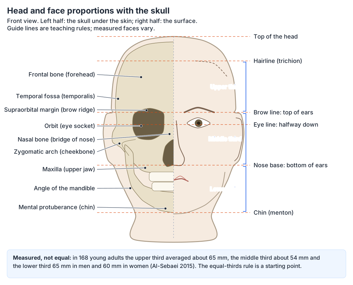
The classic teaching rules for the front of the head: the eyes are halfway between the top of the head and the chin, the bottom of the nose is halfway between the eyes and the chin, the mouth is halfway between the nose and the chin, the corners of the mouth line up with the centres of the eyes, and the ears run from the eyebrows down to the bottom of the nose. The same drawing lab adds that all these ideal measurements vary from face to face. (University of Evansville Figure Drawing Lab: Face proportions.)
Measured faces confirm the warning. In 168 young adults the face was not divided into three equal thirds: the upper third (hairline to nasion) averaged about 65 mm, the middle third (nasion to the base of the nose) about 54 mm and the lower third about 65 mm in men and 60 mm in women. About 91 percent of the men and 88 percent of the women had a longer lower third than middle third, and most had a gap between the eyes wider than one eye's width. (Al-Sebaei MO. The validity of three neo-classical facial canons in young adults originating from the Arabian Peninsula. Head Face Med. 2015;11:4.)
The skull gives the face its planes. The frontal bone forms the forehead, the supraorbital margin is the upper rim of each eye socket, the paired nasal bones form the bridge of the nose and the zygomatic arch runs from the cheek to just above the ear canal. The mandible, the only movable bone of the skull, has a horizontal body and a vertical ramus that meet at the angle of the jaw, and its forward projection forms the chin. (OpenStax Anatomy and Physiology 2e, 7.2: The Skull.)
Most facial muscles insert into the skin rather than bone, which is how they move it into expressions. The circular orbicularis oculi closes the eye, orbicularis oris shapes the lips, occipitofrontalis raises the scalp and eyebrows, the masseter is the main muscle used for chewing, and the temporalis can be felt at the temple. (OpenStax Anatomy and Physiology 2e, 11.3: Axial Muscles of the Head, Neck, and Back.)
Every muscle of facial expression, with its action and nerve, is labelled in the guide to the Facial muscles.
Hand anatomy drawing: bones, knuckles and size
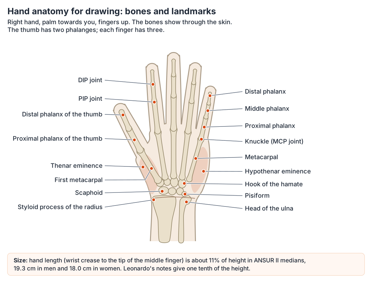
The wrist has eight carpal bones in two rows (scaphoid, lunate, triquetrum and pisiform; trapezium, trapezoid, capitate and hamate), the palm five metacarpals, and the digits 14 phalanges: two in the thumb and three in each finger. The knuckles at the base of the fingers are the rounded distal ends of the metacarpals. (OpenStax Anatomy and Physiology 2e, 8.2: Bones of the Upper Limb.)
Hand length, from the wrist crease to the tip of the middle finger, is about 11 percent of height in the ANSUR II medians: 19.3 cm in men and 18.0 cm in women. The elbow-to-fingertip length is about 27 percent of height. (The Ergonomics Center, NC State University: Anthropometric summary tables (ANSUR II, Gordon et al. 2014).)
The canons are close but not identical. Leonardo's notes give the whole hand as one tenth of a man's height, the same as the face from the hairline to the chin, and the elbow to the tip of the hand as one fifth. (University of Evansville Figure Drawing Lab: Vitruvian proportions.)
The Evansville drawing lab gives the hand, from the wrist to the outstretched fingertips, as one head long and the elbow to the fingertips as two heads. Compared with the measured averages cited above (hand about 11 percent of height, head about 13 percent in Mather's soldiers), the real hand is a little shorter than the head. (University of Evansville Figure Drawing Lab: Body proportions.)
The bones, joints, tendons and muscles of the hand are covered in detail in the guide to Hand anatomy.
Using the 3D model as an anatomy pose and structure reference
The 3D model at the top of the page comes from BodyParts3D, a database of 3D anatomical structures built on a whole-body model of an adult human male and released under a CC BY licence. (BodyParts3D, Database Center for Life Science.)
It opens with the landmark bones and the figure-shaping muscles highlighted. Drag to rotate the body to any angle, use the Front, Back, Left, Right and Top buttons for exact views, slide the Muscles control down to see the skeleton and its landmarks through the muscles, and switch on Hover labels to name each part. Rotating to three-quarter and top views is a quick way to study how the rib cage, pelvis and limbs foreshorten.
Proportion systems have a known limit: they work only when the figure is not foreshortened. That is where a rotatable model helps, because it shows the same body from every angle. (Proko: Human Proportions, Average Figure (lesson notes).)
Be aware of the model's limits. It stands in one pose and its joints cannot be bent, so it is a structure reference rather than a pose library. It has no rectus abdominis, latissimus dorsi, chewing muscles or muscles of facial expression apart from the platysma, so use the diagrams here for those. The Female switch shows the female reproductive organs, pelvis and breasts, but the female body shares the male skeleton and muscles.
The full muscular system in 3D, with every modelled muscle, is in the guide to the 3D muscle model.
Anatomy drawing practice: tips that use the anatomy
Study real people before stylising. One useful exercise is to draw the same character at realistic, moderate and extreme proportions to see what can change and what must stay. Practise forms in perspective and foreshortening, simplify the hand into palm, thumb and grouped fingers and the foot into a wedge, and learn the major muscle groups that affect the silhouette before the small ones. (The Artillerist: Anatomy for Concept Artists: What You Actually Need to Learn.)
Use the landmarks to build the pose. Connecting the pit of the neck, the lower end of the sternum, the navel and the pubic bone with a C or S curve gives the gesture of the torso, which usually drives the rest of the pose. At the elbow, the two epicondyles and the olecranon line up when the arm is straight and form a triangle when it bends. (Proko: Landmarks of the Human Body.)
Treat every canon as a starting point. Richer's system is meant as a reference to measure from, and George Bridgman, quoted in the same lesson, warned that drawing the ideal average produces a drawing without character. (Proko: Human Proportions, Average Figure (lesson notes).)
The anatomical position and the terms anatomists use for directions are explained in Directional terms.
Anatomy drawing reference in the 3D model
- Abdominal part of pectoralis major
- The lowest fibres of pectoralis major, arising from the abdominal wall.
- Acromial part of deltoid
- The middle fibres of the deltoid, forming the rounded contour of the shoulder.
- Ascending part of trapezius
- The lower fibres of trapezius, running upward and laterally to the scapular spine.
- Body of sternum
- The long middle part of the sternum.
- Brachioradialis
- An elbow flexor that forms the lateral border of the cubital fossa; unusually for a flexor, it is supplied by the radial nerve.
- Calcaneal tendon
- The thick tendon joining gastrocnemius and soleus to the heel bone.
- Calcaneus
- The largest tarsal bone, forming the heel.
- Clavicle
- The collarbone, an S-shaped long bone running between the sternum and the shoulder.
- Clavicular part of deltoid
- The anterior fibres of the deltoid.
- Clavicular part of pectoralis major
- The upper fibres of pectoralis major.
- Descending part of trapezius
- The upper fibres of trapezius, running down from the skull and neck.
- External oblique
- The largest and most superficial lateral abdominal wall muscle.
- Femur
- The thigh bone, the longest, heaviest and strongest bone in the body.
- Fibula
- The slender lateral bone of the leg.
- Fibularis longus
- A long lateral leg muscle whose tendon crosses the sole of the foot.
- Frontal bone
- The bone of the forehead and the roof of the orbits.
- Gluteus maximus
- The largest and most superficial buttock muscle.
- Gluteus medius
- A fan-shaped muscle under gluteus maximus on the outer ilium.
- Hip bone
- The large irregular bone of the pelvis formed by the fusion of the ilium, ischium and pubis.
- Humerus
- The long bone of the upper arm.
- Iliotibial tract
- A thick band of fascia lata on the outer thigh.
- Lateral head of gastrocnemius
- The lateral head of the superficial calf muscle.
- Lateral head of triceps brachii
- The lateral head of the triceps.
- Long head of biceps brachii
- The long head of biceps, passing through the shoulder joint.
- Long head of biceps femoris
- The long, superficial head of biceps femoris, the lateral hamstring.
- Long head of triceps brachii
- The long head of triceps, crossing the shoulder joint.
- Mandible
- The lower jaw bone.
- Manubrium
- The upper part of the sternum.
- Medial head of gastrocnemius
- The larger medial head of the superficial calf muscle.
- Patella
- The kneecap, a triangular sesamoid bone in the tendon of quadriceps femoris.
- Radius
- The lateral bone of the forearm.
- Rectus femoris
- The only part of quadriceps femoris that crosses both the hip and knee.
- Sacrum
- A triangular bone of five fused sacral vertebrae.
- Sartorius
- The longest muscle in the body, a strap-like muscle crossing the thigh obliquely.
- Scapula
- The shoulder blade, a flat triangular bone on the back of the thorax.
- Semitendinosus
- A hamstring muscle with a long cord-like tendon on the posteromedial thigh.
- Serratus anterior
- A broad muscle on the side of the thorax that holds the scapula against the chest wall.
- Seventh cervical vertebra
- The seventh of the seven cervical (neck) vertebrae, labelled C7.
- Short head of biceps brachii
- The medial head of biceps brachii at the front of the arm.
- Soleus
- A broad flat calf muscle lying deep to gastrocnemius.
- Spinal part of deltoid
- The posterior fibers of deltoid arising from the scapular spine.
- Sternocleidomastoid
- A thick superficial neck muscle forming the main landmark dividing the anterior and posterior triangles.
- Sternocostal part of pectoralis major
- The large lower part of pectoralis major arising from the sternum and ribs.
- Tensor fasciae latae
- A small hip muscle at the front and side of the thigh.
- Tibia
- The shinbone, the medial and main weight-bearing bone of the leg.
- Tibialis anterior
- The main dorsiflexor on the front of the leg.
- Transverse part of trapezius
- The middle fibers of trapezius, running horizontally.
- Ulna
- The medial bone of the forearm.
- Vastus lateralis
- The largest quadriceps muscle on the lateral thigh.
- Vastus medialis
- The medial quadriceps muscle with oblique fibers near the patella.
- Xiphoid process
- The small lowest part of the sternum.
- Zygomatic bone
- The cheekbone.
Frequently asked questions
How many heads tall is a person when drawing?
Most artists use 7½ heads for an average adult, from Paul Richer's canon, and 8 heads for an idealised or heroic figure, as in Leonardo's Vitruvian Man. Measured soldiers averaged about 7.7 heads, with individuals ranging from about 6½ to 10. Some drawing teachers say most people are 6 to 7 heads, so treat any canon as a starting point.
What are the main landmarks of the body for drawing?
The most useful are bony points close to the skin: the pit of the neck, clavicles, acromion at the tip of the shoulder, sternum, the elbow points, the wrist bones, the iliac crest and ASIS at the hip, the greater trochanter, the kneecap and the ankle bones. On the back, use the C7 vertebra, the scapula and the two dimples over the PSIS.
How do I start learning anatomy for drawing?
Start with gesture, then learn the skeleton as a framework: skull, spine, rib cage, pelvis and limb bones. Next learn the bony landmarks you can see at the surface, then the large muscle groups that change the silhouette, such as the deltoid, pectoralis major, latissimus dorsi, gluteus maximus and quadriceps. Practise with reference from several angles, including a rotatable 3D model.
How do male and female proportions differ in drawing?
In measured medians, women are about 7 percent shorter, with shoulders about 12 percent narrower across the deltoids but hips slightly wider in absolute terms. The female pelvis is wider with a larger subpubic angle, the carrying angle at the elbow is larger, and women tend to store more fat at the hips and thighs. Individual variation is far larger than these averages.
Where do the eyes go on a drawn face?
The usual rule is that the eyes sit halfway between the top of the head and the chin, which surprises beginners who place them too high. The bottom of the nose is about halfway between the eyes and the chin, and the ears run from the eyebrows to the bottom of the nose. Measured faces vary, so check the rules against your model.
How big is the hand compared with the face?
In Leonardo's canon the hand and the face from the hairline to the chin are both one tenth of the height, so they are the same length. Measured hands are about 11 percent of height, 19.3 cm in men and 18.0 cm in women, which is a little shorter than the full head height from crown to chin.
Can I use a 3D model as an anatomy pose reference?
A rotatable 3D model is an excellent structure reference: you can turn the body to any angle, see how the rib cage and pelvis foreshorten, and see the skeleton under the muscles. The model on this page stands in one pose and cannot be bent, so pair it with photographs or life drawing when you need a specific pose.
References
- Mather G. Head-body ratio as a visual cue for stature in people and sculptural art. Perception. 2010;39(10):1390-1395. doi:10.1068/p6737
- Proko. Human Proportions: Average Figure (Paul Richer's canon), lesson notes.
- Proko. Landmarks of the Human Body, lesson.
- Larmann R. Figure Drawing Lab: Body proportions. University of Evansville.
- Larmann R. Figure Drawing Lab: Face proportions. University of Evansville.
- Larmann R. Figure Drawing Lab: Vitruvian proportions. University of Evansville.
- The Ergonomics Center, NC State University. Anthropometric Data for U.S. Adults, from Gordon CC et al. 2012 Anthropometric Survey of U.S. Army Personnel: Methods and Summary Statistics, 2014.
- Penn State OpenLab. ANSUR II: anthropometric survey data.
- Centers for Disease Control and Prevention, National Center for Health Statistics. FastStats: Body Measurements.
- Al-Sebaei MO. The validity of three neo-classical facial canons in young adults originating from the Arabian Peninsula. Head Face Med. 2015;11:4. doi:10.1186/s13005-015-0064-y
- Lee MJ, Fried SK. Sex-dependent depot differences in adipose tissue development and function; role of sex steroids. J Obes Metab Syndr. 2017;26:172-180.
- Wight LC. How to Draw Bodies with Landmarks to Anatomy. Artists Network.
- The Artillerist. Anatomy for Concept Artists: What You Actually Need to Learn.
- OpenStax. Anatomy and Physiology 2e, §7.2 The Skull. Rice University, 2022.
- OpenStax. Anatomy and Physiology 2e, §7.3 The Vertebral Column. Rice University, 2022.
- OpenStax. Anatomy and Physiology 2e, §7.4 The Thoracic Cage. Rice University, 2022.
- OpenStax. Anatomy and Physiology 2e, §8.1 The Pectoral Girdle. Rice University, 2022.
- OpenStax. Anatomy and Physiology 2e, §8.2 Bones of the Upper Limb. Rice University, 2022.
- OpenStax. Anatomy and Physiology 2e, §8.3 The Pelvic Girdle and Pelvis. Rice University, 2022.
- OpenStax. Anatomy and Physiology 2e, §8.4 Bones of the Lower Limb. Rice University, 2022.
- OpenStax. Anatomy and Physiology 2e, §11.3 Axial Muscles of the Head, Neck, and Back. Rice University, 2022.
- OpenStax. Anatomy and Physiology 2e, §11.4 Axial Muscles of the Abdominal Wall and Thorax. Rice University, 2022.
- OpenStax. Anatomy and Physiology 2e, §11.5 Muscles of the Pectoral Girdle and Upper Limbs. Rice University, 2022.
- OpenStax. Anatomy and Physiology 2e, §11.6 Appendicular Muscles of the Pelvic Girdle and Lower Limbs. Rice University, 2022.
- Texas Tech University Health Sciences Center El Paso. Medical Gross Anatomy: Topographical anatomy of the thorax.
- Texas Tech University Health Sciences Center El Paso. Medical Gross Anatomy: Topographical anatomy of the abdomen.
- Texas Tech University Health Sciences Center El Paso. Medical Gross Anatomy: Topographical anatomy of the back.
- Breast. Radiopaedia reference article.
- BodyParts3D. Database Center for Life Science, Japan.
- Mitsuhashi N. et al. (2009). BodyParts3D: 3D structure database for anatomical concepts. Nucleic Acids Research 37, D782–D785. Data © The Database Center for Life Science, CC BY 4.0.
Related anatomy
- Human body diagramHuman anatomy diagram set: eight labelled drawings of body parts, internal organs, skeleton and muscles front and…
- Body regionsRegional terms in anatomy with front and back body-region diagrams: cephalic, cervical, brachial, femoral, crural and…
- Skeletal systemExplore a realistic 3D human skeleton: the 206 bones, axial vs appendicular skeleton, bone types, bony landmarks and…
- Muscular systemExplore an interactive 3D muscle model: how muscles move bones, how they are named, the upper body and leg muscles…
- Female anatomyFemale anatomy explained with a labelled body diagram and 3D model: reproductive organs, vulva, breasts, pelvis…
- Facial musclesFace muscles labeled: labelled diagrams and a table of every muscle of facial expression, from procerus and…
Explore the whole body in the interactive 3D human anatomy model, or browse the A–Z list of human structures.